 Movie
Movie Controller

[English] 日本語
 Yorodumi
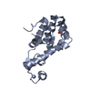
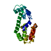
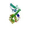
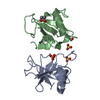
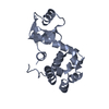
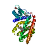
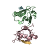
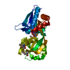
- PDB-3jr6: Sequential reorganization of beta-sheet topology by insertion of ... -
+ Open data

- Basic information
| Entry | Database: PDB / ID: 3jr6 | |||||||||
|---|---|---|---|---|---|---|---|---|---|---|
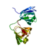
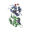
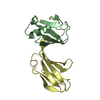
| Title | Sequential reorganization of beta-sheet topology by insertion of a single strand | |||||||||
 Components Components | Lysozyme | |||||||||
 Keywords Keywords | HYDROLASE / sequence duplication / protein design / tandem repeat / beta-sheet / Antimicrobial / Bacteriolytic enzyme / Glycosidase | |||||||||
| Function / homology |  Function and homology information Function and homology informationviral release from host cell by cytolysis / peptidoglycan catabolic process / cell wall macromolecule catabolic process / lysozyme / lysozyme activity / host cell cytoplasm / defense response to bacterium Similarity search - Function | |||||||||
| Biological species |  Enterobacteria phage T4 (virus) Enterobacteria phage T4 (virus) | |||||||||
| Method |  X-RAY DIFFRACTION / MOLECULAR REPLACEMENT / Resolution: 3 Å X-RAY DIFFRACTION / MOLECULAR REPLACEMENT / Resolution: 3 Å | |||||||||
 Authors Authors | Sagermann, M. / Baas, W.A. / Matthews, B.W. | |||||||||
 Citation Citation | Journal: Protein Sci. / Year: 2006 Title: Sequential reorganization of beta-sheet topology by insertion of a single strand. Authors: Sagermann, M. / Baase, W.A. / Matthews, B.W. #1: Journal: Proc.Natl.Acad.Sci.USA / Year: 2003Title: Long-distance conformational changes in a protein engineered by modulated sequence duplication. Authors: Sagermann, M. / Gay, L. / Matthews, B.W. #2: Journal: Proc.Natl.Acad.Sci.USA / Year: 1999Title: Structural characterization of an engineered tandem repeat contrasts the importance of context and sequence in protein folding. Authors: Sagermann, M. / Baase, W.A. / Matthews, B.W. | |||||||||
| History |
|
- Structure visualization
| Structure viewer | Molecule: MolmilJmol/JSmol |
|---|
- Downloads & links
-Download
| PDBx/mmCIF format | 3jr6.cif.gz | 128.9 KB | Display | PDBx/mmCIF format |
|---|---|---|---|---|
| PDB format | pdb3jr6.ent.gz | 101.9 KB | Display | PDB format |
| PDBx/mmJSON format | 3jr6.json.gz | Tree view | PDBx/mmJSON format | |
| Others |  Other downloads Other downloads |
-Validation report
| Arichive directory | https://data.pdbj.org/pub/pdb/validation_reports/jr/3jr6ftp://data.pdbj.org/pub/pdb/validation_reports/jr/3jr6 | HTTPS FTP |
|---|
-Related structure data


-Links
 PDBj
PDBj


- Assembly
| Deposited unit | 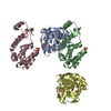
| ||||||||
|---|---|---|---|---|---|---|---|---|---|
| 1 | 
| ||||||||
| 2 | 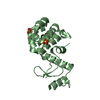
| ||||||||
| 3 | 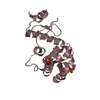
| ||||||||
| 4 | 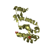
| ||||||||
| Unit cell |
|
-Components
| #1: Protein | Mass: 19220.088 Da / Num. of mol.: 4 Mutation: C54T, C97A, insertion of seuqence (gighll) after residue L33 Source method: isolated from a genetically manipulated source Source: (gene. exp.) Enterobacteria phage T4 (virus) / Gene: E / Plasmid: phs 1403 / Production host:  #2: Chemical | ChemComp-SO4 /   Mass: 96.063 Da / Num. of mol.: 8 / Source method: obtained synthetically / Formula: SO4 Mass: 96.063 Da / Num. of mol.: 8 / Source method: obtained synthetically / Formula: SO4 |
|---|
-Experimental details
-Experiment
| Experiment | Method: X-RAY DIFFRACTION / Number of used crystals: 1 |
|---|
- Sample preparation
| Crystal | Density Matthews: 2.38 Å3/Da / Density % sol: 48.29 % |
|---|---|
| Crystal grow | Temperature: 298 K / Method: vapor diffusion, hanging drop / pH: 7.5 Details: 20% PEG 3400, 50 mM ammonium sulfate, Tris, pH 7.5, VAPOR DIFFUSION, HANGING DROP, temperature 298K |
-Data collection
| Diffraction | Mean temperature: 110 K |
|---|---|
| Diffraction source | Source: ROTATING ANODE / Type: RIGAKU RU200 |
| Detector | Type: RIGAKU RAXIS II / Detector: IMAGE PLATE / Date: Sep 5, 1998 / Details: yale mirrors |
| Radiation | Protocol: SINGLE WAVELENGTH / Monochromatic (M) / Laue (L): M / Scattering type: x-ray |
| Radiation wavelength | Relative weight: 1 |
| Reflection | Resolution: 3→32.44 Å / Num. obs: 17925 / % possible obs: 96.5 % / Observed criterion σ(F): 0 / Observed criterion σ(I): 0 / Redundancy: 4.3 % / Rsym value: 0.07 |
| Reflection shell | Highest resolution: 3 Å / Rsym value: 0.097 / % possible all: 99.8 |
- Processing
| Software |
| |||||||||||||||||||||||||
|---|---|---|---|---|---|---|---|---|---|---|---|---|---|---|---|---|---|---|---|---|---|---|---|---|---|---|
| Refinement | Method to determine structure: MOLECULAR REPLACEMENT Starting model: T4 lysozyme I3P mutant Resolution: 3→32.44 Å / Cross valid method: THROUGHOUT / σ(F): 0 / σ(I): 0 / Stereochemistry target values: Engh & Huber Details: Authors state that scaling and B-value refinement were performed with CNS using a recommended resolution range of 6.0-3.0 A resolution. As the B-values for the data set were determined to be ...Details: Authors state that scaling and B-value refinement were performed with CNS using a recommended resolution range of 6.0-3.0 A resolution. As the B-values for the data set were determined to be low (even for higher low-resolution cut-offs), the B-values for the atoms remained similarly low as well. Molecules A and B exhibit well-defined density of the loops. The corresponding loops of molecules B and C were in poor density but could clearly be identified. Loops and terminal residues that could not be refined reliably were omitted: B(L170), D(L170), B(34-46), C(36-46).
| |||||||||||||||||||||||||
| Displacement parameters |
| |||||||||||||||||||||||||
| Refinement step | Cycle: LAST / Resolution: 3→32.44 Å
| |||||||||||||||||||||||||
| Refine LS restraints |
|
