 Movie
Movie Controller

[English] 日本語
 Yorodumi
- PDB-261l: STRUCTURAL CHARACTERISATION OF AN ENGINEERED TANDEM REPEAT CONTRA... -
+ Open data

- Basic information
| Entry | Database: PDB / ID: 261l | ||||||
|---|---|---|---|---|---|---|---|
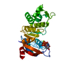
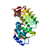
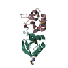
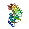
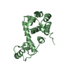
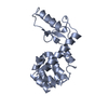
| Title | STRUCTURAL CHARACTERISATION OF AN ENGINEERED TANDEM REPEAT CONTRASTS THE IMPORTANCE OF CONTEXT AND SEQUENCE IN PROTEIN FOLDING | ||||||
 Components Components | LYSOZYME | ||||||
 Keywords Keywords | HYDROLASE / HYDROLASE (O-GLYCOSYL) / T4 LYSOZYME / ENGINEERED TANDEM REPEAT / PROTEIN ENGINEERING / PROTEIN DESIGN | ||||||
| Function / homology |  Function and homology information Function and homology informationviral release from host cell by cytolysis / peptidoglycan catabolic process / cell wall macromolecule catabolic process / lysozyme / lysozyme activity / host cell cytoplasm / defense response to bacterium Similarity search - Function | ||||||
| Biological species |  Enterobacteria phage T4 (virus) Enterobacteria phage T4 (virus) | ||||||
| Method |  X-RAY DIFFRACTION / SYNCHROTRON / MOLECULAR REPLACEMENT / Resolution: 2.5 Å X-RAY DIFFRACTION / SYNCHROTRON / MOLECULAR REPLACEMENT / Resolution: 2.5 Å | ||||||
 Authors Authors | Sagermann, M. / Baase, W.A. / Matthews, B.W. | ||||||
 Citation Citation | Journal: Proc.Natl.Acad.Sci.USA / Year: 1999 Title: Structural characterization of an engineered tandem repeat contrasts the importance of context and sequence in protein folding. Authors: Sagermann, M. / Baase, W.A. / Matthews, B.W. #1: Journal: Protein Sci. / Year: 1998Title: Protein Structural Plasticity Examplified by Insertion and Deletion Authors: Vetter, I.R. / Baase, W.A. / Heinz, D. / Xiong, J.P. / Snow, S. / Matthews, B.W. #2: Journal: Nature / Year: 1993Title: Folding and Function of a T4 Lysosyme Containing 10 Consecutive Alanines Illustrate the Redundancy of Information in an Amino Acid Sequence Authors: Heinz, D. / Baase, W.A. / Dahlquist, F.W. / Matthews, B.W. | ||||||
| History |
|
- Structure visualization
| Structure viewer | Molecule: MolmilJmol/JSmol |
|---|
- Downloads & links
-Download
| PDBx/mmCIF format | 261l.cif.gz | 47 KB | Display | PDBx/mmCIF format |
|---|---|---|---|---|
| PDB format | pdb261l.ent.gz | 33.3 KB | Display | PDB format |
| PDBx/mmJSON format | 261l.json.gz | Tree view | PDBx/mmJSON format | |
| Others |  Other downloads Other downloads |
-Validation report
| Arichive directory | https://data.pdbj.org/pub/pdb/validation_reports/61/261lftp://data.pdbj.org/pub/pdb/validation_reports/61/261l | HTTPS FTP |
|---|
-Related structure data

-Links
 PDBj
PDBj


- Assembly
| Deposited unit | 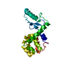
| ||||||||
|---|---|---|---|---|---|---|---|---|---|
| 1 |
| ||||||||
| Unit cell |
|
-Components
| #1: Protein | Mass: 19544.395 Da / Num. of mol.: 1 / Mutation: L39I Source method: isolated from a genetically manipulated source Source: (gene. exp.) Enterobacteria phage T4 (virus) / Genus: T4-like viruses / Species: Enterobacteria phage T4 sensu latoDescription: BACTERIOPHAGE T4 (MUTANT GENE DERIVED FROM THE M13 PLASMID BY CLONING THE T4 LY SOZYME GENE) Cellular location: CYTOPLASM / Gene: GENE E FROM BACTERIOPHAGE T4 / Plasmid: PHS1403 / Gene (production host): T4 LYSOZYME / Production host:  |
|---|---|
| #2: Water | ChemComp-HOH /  Mass: 18.015 Da / Num. of mol.: 43 / Source method: isolated from a natural source / Formula: H2O Mass: 18.015 Da / Num. of mol.: 43 / Source method: isolated from a natural source / Formula: H2O |
-Experimental details
-Experiment
| Experiment | Method: X-RAY DIFFRACTION / Number of used crystals: 1 |
|---|
- Sample preparation
| Crystal | Density Matthews: 2.98 Å3/Da / Density % sol: 58 % | |||||||||||||||||||||||||
|---|---|---|---|---|---|---|---|---|---|---|---|---|---|---|---|---|---|---|---|---|---|---|---|---|---|---|
| Crystal grow | pH: 7.5 Details: CRYSTALS WERE GROWN FROM 20% POLYETHYLENE GLYCOL 6000, 20% ISOPROPANOL, 50MM TRIS-HCL PH 7.5 | |||||||||||||||||||||||||
| Crystal | *PLUS | |||||||||||||||||||||||||
| Crystal grow | *PLUS Method: unknown | |||||||||||||||||||||||||
| Components of the solutions | *PLUS
|
-Data collection
| Diffraction | Ambient pressure: 101 kPa / Mean temperature: 298 K |
|---|---|
| Diffraction source | Source: SYNCHROTRON / Site: CHESS  / Beamline: A1 / Wavelength: 0.908 / Beamline: A1 / Wavelength: 0.908 |
| Detector | Type: ADSC QUANTUM 1 / Detector: CCD |
| Radiation | Protocol: SINGLE WAVELENGTH / Monochromatic (M) / Laue (L): M / Scattering type: x-ray / Wavelength: 0.908 Å |
| Radiation wavelength | Wavelength: 0.908 Å / Relative weight: 1 |
| Reflection | Resolution: 2.5→30 Å / Num. obs: 28161 / % possible obs: 86.5 % / Redundancy: 4.3 % / Biso Wilson estimate: 37.5 Å2 / Rmerge(I) obs: 0.084 |
| Reflection shell | Highest resolution: 2.5 Å / Rmerge(I) obs: 0.2 / Mean I/σ(I) obs: 2 |
- Processing
| Software |
| ||||||||||||||||||||||||||||||
|---|---|---|---|---|---|---|---|---|---|---|---|---|---|---|---|---|---|---|---|---|---|---|---|---|---|---|---|---|---|---|---|
| Refinement | Method to determine structure: MOLECULAR REPLACEMENT Starting model: CYS-FREE VERSION OF T4-LYSOZYME Resolution: 2.5→30 Å / σ(F): 0 / Stereochemistry target values: TNT PROTGEO Details: GROUPED OCCUPANCY REFINEMENT ONLY FOR RESIDUES 36-40
| ||||||||||||||||||||||||||||||
| Solvent computation | Solvent model: BABENET SCALING / Bsol: 150 Å2 / ksol: 0.7 e/Å3 | ||||||||||||||||||||||||||||||
| Refinement step | Cycle: LAST / Resolution: 2.5→30 Å
| ||||||||||||||||||||||||||||||
| Refine LS restraints |
| ||||||||||||||||||||||||||||||
| Software | *PLUS Name: TNT / Version: 5F / Classification: refinement | ||||||||||||||||||||||||||||||
| Refinement | *PLUS Rfactor obs: 0.17 / Rfactor Rwork: 0.17 | ||||||||||||||||||||||||||||||
| Solvent computation | *PLUS | ||||||||||||||||||||||||||||||
| Displacement parameters | *PLUS | ||||||||||||||||||||||||||||||
| Refine LS restraints | *PLUS
|
