 Movie
Movie Controller

[English] 日本語
 Yorodumi
- PDB-2f2q: High resolution crystal structure of T4 lysozyme mutant L20R63/A ... -
+ Open data

- Basic information
| Entry | Database: PDB / ID: 2f2q | ||||||
|---|---|---|---|---|---|---|---|
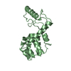
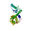
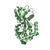
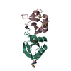
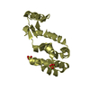
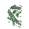
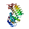
| Title | High resolution crystal structure of T4 lysozyme mutant L20R63/A liganded to guanidinium ion | ||||||
 Components Components | Lysozyme | ||||||
 Keywords Keywords | HYDROLASE / MOLECULAR SWITCH / T4 LYSOZYME / NANO-BITECHNOLOGY / PROTEIN ENGINEERING / PROTEIN DESIGN | ||||||
| Function / homology |  Function and homology information Function and homology informationviral release from host cell by cytolysis / peptidoglycan catabolic process / cell wall macromolecule catabolic process / lysozyme / lysozyme activity / host cell cytoplasm / defense response to bacterium Similarity search - Function | ||||||
| Biological species |  Enterobacteria phage T4 (virus) Enterobacteria phage T4 (virus) | ||||||
| Method |  X-RAY DIFFRACTION / SYNCHROTRON / MOLECULAR REPLACEMENT / Resolution: 1.45 Å X-RAY DIFFRACTION / SYNCHROTRON / MOLECULAR REPLACEMENT / Resolution: 1.45 Å | ||||||
 Authors Authors | Yousef, M.S. / Bischoff, N. / Dyer, C.M. / Baase, W.A. / Matthews, B.W. | ||||||
 Citation Citation | Journal: Protein Sci. / Year: 2006 Title: Guanidinium derivatives bind preferentially and trigger long-distance conformational changes in an engineered T4 lysozyme. Authors: Yousef, M.S. / Bischoff, N. / Dyer, C.M. / Baase, W.A. / Matthews, B.W. #1: Journal: Proc.Natl.Acad.Sci.USA / Year: 2004Title: Use of sequence duplication to engineer a ligand-triggered, long-distance molecular switch in T4 lysozyme Authors: Yousef, M.S. / Baase, W.A. / Matthews, B.W. #2: Journal: Proc.Natl.Acad.Sci.USA / Year: 1999Title: Structural characterization of an engineered tandem repeat contrasts the importance of context and sequence in protein folding Authors: Sagermann, M. / Baase, W.A. / Matthews, B.W. #3: Journal: Proc.Natl.Acad.Sci.USA / Year: 2003Title: Long-distance conformational changes in a protein engineered by modulated sequence duplication Authors: Sagermann, M. / Gay, L. / Matthews, B.W. | ||||||
| History |
|
- Structure visualization
| Structure viewer | Molecule: MolmilJmol/JSmol |
|---|
- Downloads & links
-Download
| PDBx/mmCIF format | 2f2q.cif.gz | 53.8 KB | Display | PDBx/mmCIF format |
|---|---|---|---|---|
| PDB format | pdb2f2q.ent.gz | 37.4 KB | Display | PDB format |
| PDBx/mmJSON format | 2f2q.json.gz | Tree view | PDBx/mmJSON format | |
| Others |  Other downloads Other downloads |
-Validation report
| Arichive directory | https://data.pdbj.org/pub/pdb/validation_reports/f2/2f2qftp://data.pdbj.org/pub/pdb/validation_reports/f2/2f2q | HTTPS FTP |
|---|
-Related structure data
| Related structure data | 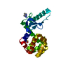 2f32C  2f47C 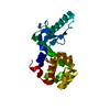 1t8aS S: Starting model for refinement C: citing same article ( |
|---|---|
| Similar structure data |


-Links
 PDBj
PDBj


- Assembly
| Deposited unit | 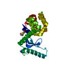
| ||||||||
|---|---|---|---|---|---|---|---|---|---|
| 1 |
| ||||||||
| Unit cell |
|
-Components
| #1: Protein | Mass: 19685.541 Da / Num. of mol.: 1 / Mutation: I39L, A63R, T65C, A108C Source method: isolated from a genetically manipulated source Source: (gene. exp.) Enterobacteria phage T4 (virus) / Genus: T4-like viruses / Species: Enterobacteria phage T4 sensu lato / Gene: E / Plasmid: pHS1403 / Production host:  |
|---|---|
| #2: Chemical | ChemComp-CL /   Mass: 35.453 Da / Num. of mol.: 1 / Source method: obtained synthetically / Formula: Cl Mass: 35.453 Da / Num. of mol.: 1 / Source method: obtained synthetically / Formula: Cl |
| #3: Chemical | ChemComp-GAI /   Mass: 59.070 Da / Num. of mol.: 1 / Source method: obtained synthetically / Formula: CH5N3 Mass: 59.070 Da / Num. of mol.: 1 / Source method: obtained synthetically / Formula: CH5N3 |
| #4: Chemical | ChemComp-HED /   Mass: 154.251 Da / Num. of mol.: 1 / Source method: obtained synthetically / Formula: C4H10O2S2 Mass: 154.251 Da / Num. of mol.: 1 / Source method: obtained synthetically / Formula: C4H10O2S2 |
| #5: Water | ChemComp-HOH /  Mass: 18.015 Da / Num. of mol.: 181 / Source method: isolated from a natural source / Formula: H2O Mass: 18.015 Da / Num. of mol.: 181 / Source method: isolated from a natural source / Formula: H2O |
-Experimental details
-Experiment
| Experiment | Method: X-RAY DIFFRACTION / Number of used crystals: 1 |
|---|
- Sample preparation
| Crystal | Density Matthews: 2.64 Å3/Da / Density % sol: 53 % |
|---|---|
| Crystal grow | Temperature: 277 K / pH: 6.5 Details: 0.5 mM protein, 1.8 M MIXED POTASSIUM AND SODIUM PHOSPHATE, 0.2 M GUANIDINIUM CHLORIDE, PH 6.5, VAPOR DIFFUSION, HANGING DROP, TEMPERATURE 277K, pH 6.50 |
-Data collection
| Diffraction | Mean temperature: 100 K |
|---|---|
| Diffraction source | Source: SYNCHROTRON / Site: ALS  / Beamline: 8.2.1 / Wavelength: 0.953 / Beamline: 8.2.1 / Wavelength: 0.953 |
| Detector | Type: ADSC / Detector: CCD / Date: May 26, 2005 / Details: KOHZU: Double Crystal Si(111) |
| Radiation | Monochromator: Double crystal, Si(111 / Protocol: SINGLE WAVELENGTH / Monochromatic (M) / Laue (L): M / Scattering type: x-ray |
| Radiation wavelength | Wavelength: 0.953 Å / Relative weight: 1 |
| Reflection | Resolution: 1.45→30 Å / Num. obs: 36743 / % possible obs: 90 % / Biso Wilson estimate: 18.7 Å2 / Rsym value: 0.077 / Net I/σ(I): 27 |
| Reflection shell | Resolution: 1.45→1.5 Å / Mean I/σ(I) obs: 3.6 / Rsym value: 0.228 / % possible all: 89 |
- Processing
| Software |
| ||||||||||||||||||||||||||||||||||||||||||||||||||||||||||||||||||||||||||||||||
|---|---|---|---|---|---|---|---|---|---|---|---|---|---|---|---|---|---|---|---|---|---|---|---|---|---|---|---|---|---|---|---|---|---|---|---|---|---|---|---|---|---|---|---|---|---|---|---|---|---|---|---|---|---|---|---|---|---|---|---|---|---|---|---|---|---|---|---|---|---|---|---|---|---|---|---|---|---|---|---|---|---|
| Refinement | Method to determine structure: MOLECULAR REPLACEMENT Starting model: PDB ENTRY 1T8A Resolution: 1.45→30 Å / Rfactor Rfree error: 0.006 / Data cutoff high absF: 1714737.73 / Data cutoff low absF: 0 / Isotropic thermal model: RESTRAINED / Cross valid method: THROUGHOUT / σ(F): 0
| ||||||||||||||||||||||||||||||||||||||||||||||||||||||||||||||||||||||||||||||||
| Solvent computation | Solvent model: FLAT MODEL / Bsol: 49.1 Å2 / ksol: 0.39 e/Å3 | ||||||||||||||||||||||||||||||||||||||||||||||||||||||||||||||||||||||||||||||||
| Displacement parameters | Biso mean: 22.4 Å2
| ||||||||||||||||||||||||||||||||||||||||||||||||||||||||||||||||||||||||||||||||
| Refine analyze |
| ||||||||||||||||||||||||||||||||||||||||||||||||||||||||||||||||||||||||||||||||
| Refinement step | Cycle: LAST / Resolution: 1.45→30 Å
| ||||||||||||||||||||||||||||||||||||||||||||||||||||||||||||||||||||||||||||||||
| Refine LS restraints |
| ||||||||||||||||||||||||||||||||||||||||||||||||||||||||||||||||||||||||||||||||
| LS refinement shell | Resolution: 1.45→1.54 Å / Rfactor Rfree error: 0.017 / Total num. of bins used: 6
| ||||||||||||||||||||||||||||||||||||||||||||||||||||||||||||||||||||||||||||||||
| Xplor file |
|
