[English] 日本語
Yorodumi
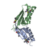
- PDB-3qht: Crystal Structure of the Monobody ySMB-1 bound to yeast SUMO -

+
Open data


ID or keywords:

Loading...

-
Basic information

Entry
Database: PDB / ID: 3qht
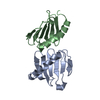
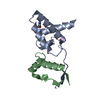
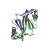
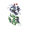
TitleCrystal Structure of the Monobody ySMB-1 bound to yeast SUMO
Components
  • Monobody ySMB-1
  • Ubiquitin-like protein SMT3
KeywordsDE NOVO PROTEIN / Fibronectin Type III / yeast small ubiquitin-like modifier / SMT3
Function / homology
Function and homology information


SUMO is conjugated to E1 (UBA2:SAE1) / SUMOylation of nuclear envelope proteins / SUMO is transferred from E1 to E2 (UBE2I, UBC9) / SUMOylation of SUMOylation proteins / SUMO is proteolytically processed / SUMOylation of transcription factors / Postmitotic nuclear pore complex (NPC) reformation / SUMOylation of transcription cofactors / septin ring / SUMOylation of DNA damage response and repair proteins ...SUMO is conjugated to E1 (UBA2:SAE1) / SUMOylation of nuclear envelope proteins / SUMO is transferred from E1 to E2 (UBE2I, UBC9) / SUMOylation of SUMOylation proteins / SUMO is proteolytically processed / SUMOylation of transcription factors / Postmitotic nuclear pore complex (NPC) reformation / SUMOylation of transcription cofactors / septin ring / SUMOylation of DNA damage response and repair proteins / SUMOylation of RNA binding proteins / SUMOylation of DNA replication proteins / SUMOylation of chromatin organization proteins / ubiquitin-like protein ligase binding / protein sumoylation / condensed nuclear chromosome / protein tag activity / identical protein binding / nucleus
Similarity search - Function
Rad60/SUMO-like domain / Ubiquitin-2 like Rad60 SUMO-like / Phosphatidylinositol 3-kinase Catalytic Subunit; Chain A, domain 1 / Ubiquitin-like (UB roll) / Ubiquitin family / Ubiquitin homologues / Ubiquitin-like domain / Ubiquitin domain profile. / Ubiquitin-like domain superfamily / Immunoglobulins ...Rad60/SUMO-like domain / Ubiquitin-2 like Rad60 SUMO-like / Phosphatidylinositol 3-kinase Catalytic Subunit; Chain A, domain 1 / Ubiquitin-like (UB roll) / Ubiquitin family / Ubiquitin homologues / Ubiquitin-like domain / Ubiquitin domain profile. / Ubiquitin-like domain superfamily / Immunoglobulins / Roll / Immunoglobulin-like / Sandwich / Mainly Beta / Alpha Beta
Similarity search - Domain/homology
Ubiquitin-like protein SMT3
Similarity search - Component
Biological speciesSaccharomyces cerevisiae (brewer's yeast)
artificial gene (others)
MethodX-RAY DIFFRACTION / SYNCHROTRON / MOLECULAR REPLACEMENT / Resolution: 2.4 Å
AuthorsKoide, S. / Gilbreth, R.N.
CitationJournal: Proc.Natl.Acad.Sci.USA / Year: 2011
Title: Isoform-specific monobody inhibitors of small ubiquitin-related modifiers engineered using structure-guided library design.
Authors: Gilbreth, R.N. / Truong, K. / Madu, I. / Koide, A. / Wojcik, J.B. / Li, N.S. / Piccirilli, J.A. / Chen, Y. / Koide, S.
History
DepositionJan 26, 2011Deposition site: RCSB / Processing site: RCSB
Revision 1.0May 11, 2011Provider: repository / Type: Initial release
Revision 1.1Jul 13, 2011Group: Version format compliance
Revision 1.2Nov 8, 2017Group: Refinement description / Category: software
Revision 1.3Sep 13, 2023Group: Data collection / Database references ...Data collection / Database references / Derived calculations / Refinement description
Category: chem_comp_atom / chem_comp_bond ...chem_comp_atom / chem_comp_bond / database_2 / pdbx_initial_refinement_model / struct_ref_seq / struct_ref_seq_dif / struct_site
Item: _database_2.pdbx_DOI / _database_2.pdbx_database_accession ..._database_2.pdbx_DOI / _database_2.pdbx_database_accession / _struct_ref_seq.db_align_beg / _struct_ref_seq.db_align_end / _struct_ref_seq_dif.details / _struct_site.pdbx_auth_asym_id / _struct_site.pdbx_auth_comp_id / _struct_site.pdbx_auth_seq_id

-
Structure visualization

Structure viewerMolecule:
MolmilJmol/JSmol

Downloads & links

-
Assembly

Deposited unit
A: Ubiquitin-like protein SMT3
B: Ubiquitin-like protein SMT3
C: Monobody ySMB-1
D: Monobody ySMB-1
hetero molecules


Theoretical massNumber of molelcules
Total (without water)43,93412
Polymers43,1984
Non-polymers7378
Water1,53185
1
A: Ubiquitin-like protein SMT3
C: Monobody ySMB-1
hetero molecules


Theoretical massNumber of molelcules
Total (without water)22,33610
Polymers21,5992
Non-polymers7378
Water362
TypeNameSymmetry operationNumber
identity operation1_555x,y,z1
Buried area2810 Å2
ΔGint-5 kcal/mol
Surface area9280 Å2
MethodPISA
2
B: Ubiquitin-like protein SMT3
D: Monobody ySMB-1


Theoretical massNumber of molelcules
Total (without water)21,5992
Polymers21,5992
Non-polymers00
Water362
TypeNameSymmetry operationNumber
identity operation1_555x,y,z1
Buried area1470 Å2
ΔGint-6 kcal/mol
Surface area8990 Å2
MethodPISA
Unit cell
Length a, b, c (Å)59.637, 175.462, 52.830
Angle α, β, γ (deg.)90.00, 90.00, 90.00
Int Tables number18
Space group name H-MP21212

-
Components

#1: Protein Ubiquitin-like protein SMT3 / yeast small ubiquitin-like modifier (SUMO)


Mass: 11204.536 Da / Num. of mol.: 2
Source method: isolated from a genetically manipulated source
Source: (gene. exp.) Saccharomyces cerevisiae (brewer's yeast)
Gene: SMT3, YDR510W, D9719.15 / Production host: Escherichia coli (E. coli) / References: UniProt: Q12306
#2: Protein Monobody ySMB-1


Mass: 10394.215 Da / Num. of mol.: 2
Source method: isolated from a genetically manipulated source
Source: (gene. exp.) artificial gene (others) / Production host: Escherichia coli (E. coli)
#3: Chemical
ChemComp-GOL / GLYCEROL / GLYCERIN / PROPANE-1,2,3-TRIOL / Glycerol


Mass: 92.094 Da / Num. of mol.: 8 / Source method: obtained synthetically / Formula: C3H8O3
#4: Water ChemComp-HOH / water / Water


Mass: 18.015 Da / Num. of mol.: 85 / Source method: isolated from a natural source / Formula: H2O

-
Experimental details

-
Experiment

ExperimentMethod: X-RAY DIFFRACTION / Number of used crystals: 1

-
Sample preparation

CrystalDensity Matthews: 3.2 Å3/Da / Density % sol: 61.55 %
Crystal growTemperature: 292 K / Method: vapor diffusion / pH: 8
Details: 14% PEG8000, 16% glycerol, pH 8.0, VAPOR DIFFUSION, temperature 292K

-
Data collection

DiffractionMean temperature: 100 K
Diffraction sourceSource: SYNCHROTRON / Site: APS / Beamline: 21-ID-F / Wavelength: 0.97872
DetectorType: MARMOSAIC 225 mm CCD / Detector: CCD / Date: Nov 18, 2009
RadiationMonochromator: C(111) / Protocol: SINGLE WAVELENGTH / Monochromatic (M) / Laue (L): M / Scattering type: x-ray
Radiation wavelengthWavelength: 0.97872 Å / Relative weight: 1
ReflectionResolution: 2.4→50 Å / Num. all: 22586 / Num. obs: 22586 / % possible obs: 100 % / Observed criterion σ(F): 0 / Observed criterion σ(I): 0 / Redundancy: 7.1 % / Rmerge(I) obs: 0.085 / Net I/σ(I): 18.9
Reflection shellResolution: 2.395→2.457 Å / Redundancy: 6.6 % / Rmerge(I) obs: 0.643 / Mean I/σ(I) obs: 2.15 / % possible all: 96.29

-
Processing

Software
NameVersionClassification
MD2data collection
MAR345data collection
MOLREPphasing
REFMAC5.2.0019refinement
HKL-2000data reduction
HKL-2000data scaling
RefinementMethod to determine structure: MOLECULAR REPLACEMENT
Starting model: PDB ENTRIES 1FNA AND 2EKE
Resolution: 2.4→20 Å / Cor.coef. Fo:Fc: 0.925 / Cor.coef. Fo:Fc free: 0.896 / SU B: 14.399 / SU ML: 0.171 / Cross valid method: THROUGHOUT / σ(F): 0 / ESU R: 0.287 / ESU R Free: 0.24 / Stereochemistry target values: MAXIMUM LIKELIHOOD
RfactorNum. reflection% reflectionSelection details
Rfree0.27158 1151 5.1 %RANDOM
Rwork0.2231 ---
all0.22558 21341 --
obs0.22558 21341 99.74 %-
Solvent computationIon probe radii: 0.8 Å / Shrinkage radii: 0.8 Å / VDW probe radii: 1.4 Å / Solvent model: BABINET MODEL WITH MASK
Displacement parametersBiso mean: 49.816 Å2
Baniso -1Baniso -2Baniso -3
1--0.67 Å20 Å20 Å2
2---0.03 Å20 Å2
3---0.7 Å2
Refinement stepCycle: LAST / Resolution: 2.4→20 Å
ProteinNucleic acidLigandSolventTotal
Num. atoms2666 0 48 85 2799
Refine LS restraints
Refine-IDTypeDev idealDev ideal targetNumber
X-RAY DIFFRACTIONr_bond_refined_d0.0170.0222777
X-RAY DIFFRACTIONr_angle_refined_deg1.8641.9783759
X-RAY DIFFRACTIONr_dihedral_angle_1_deg7.8065336
X-RAY DIFFRACTIONr_dihedral_angle_2_deg33.57523.636121
X-RAY DIFFRACTIONr_dihedral_angle_3_deg19.32315449
X-RAY DIFFRACTIONr_dihedral_angle_4_deg18.3281516
X-RAY DIFFRACTIONr_chiral_restr0.160.2413
X-RAY DIFFRACTIONr_gen_planes_refined0.0060.022084
X-RAY DIFFRACTIONr_nbd_refined0.2320.21185
X-RAY DIFFRACTIONr_nbtor_refined0.3080.21898
X-RAY DIFFRACTIONr_xyhbond_nbd_refined0.1580.2142
X-RAY DIFFRACTIONr_symmetry_vdw_refined0.270.254
X-RAY DIFFRACTIONr_symmetry_hbond_refined0.140.212
X-RAY DIFFRACTIONr_mcbond_it1.1071.51733
X-RAY DIFFRACTIONr_mcangle_it1.92322752
X-RAY DIFFRACTIONr_scbond_it2.56831203
X-RAY DIFFRACTIONr_scangle_it3.9184.51007
LS refinement shellResolution: 2.395→2.457 Å / Total num. of bins used: 20
RfactorNum. reflection% reflection
Rfree0.356 81 -
Rwork0.247 1450 -
obs--96.29 %

+
About Yorodumi

-
News

-
Feb 9, 2022. New format data for meta-information of EMDB entries

New format data for meta-information of EMDB entries

  • Version 3 of the EMDB header file is now the official format.
  • The previous official version 1.9 will be removed from the archive.

Related info.:EMDB header

External links:wwPDB to switch to version 3 of the EMDB data model

-
Aug 12, 2020. Covid-19 info

Covid-19 info

URL: https://pdbj.org/emnavi/covid19.php

New page: Covid-19 featured information page in EM Navigator.

Related info.:Covid-19 info / Mar 5, 2020. Novel coronavirus structure data

+
Mar 5, 2020. Novel coronavirus structure data

Novel coronavirus structure data

Related info.:Yorodumi Speices / Aug 12, 2020. Covid-19 info

External links:COVID-19 featured content - PDBj / Molecule of the Month (242):Coronavirus Proteases

+
Jan 31, 2019. EMDB accession codes are about to change! (news from PDBe EMDB page)

EMDB accession codes are about to change! (news from PDBe EMDB page)

  • The allocation of 4 digits for EMDB accession codes will soon come to an end. Whilst these codes will remain in use, new EMDB accession codes will include an additional digit and will expand incrementally as the available range of codes is exhausted. The current 4-digit format prefixed with “EMD-” (i.e. EMD-XXXX) will advance to a 5-digit format (i.e. EMD-XXXXX), and so on. It is currently estimated that the 4-digit codes will be depleted around Spring 2019, at which point the 5-digit format will come into force.
  • The EM Navigator/Yorodumi systems omit the EMD- prefix.

Related info.:Q: What is EMD? / ID/Accession-code notation in Yorodumi/EM Navigator

External links:EMDB Accession Codes are Changing Soon! / Contact to PDBj

+
Jul 12, 2017. Major update of PDB

Major update of PDB

  • wwPDB released updated PDB data conforming to the new PDBx/mmCIF dictionary.
  • This is a major update changing the version number from 4 to 5, and with Remediation, in which all the entries are updated.
  • In this update, many items about electron microscopy experimental information are reorganized (e.g. em_software).
  • Now, EM Navigator and Yorodumi are based on the updated data.

External links:wwPDB Remediation / Enriched Model Files Conforming to OneDep Data Standards Now Available in the PDB FTP Archive

-
Yorodumi

Thousand views of thousand structures

  • Yorodumi is a browser for structure data from EMDB, PDB, SASBDB, etc.
  • This page is also the successor to EM Navigator detail page, and also detail information page/front-end page for Omokage search.
  • The word "yorodu" (or yorozu) is an old Japanese word meaning "ten thousand". "mi" (miru) is to see.

Related info.:EMDB / PDB / SASBDB / Comparison of 3 databanks / Yorodumi Search / Aug 31, 2016. New EM Navigator & Yorodumi / Yorodumi Papers / Jmol/JSmol / Function and homology information / Changes in new EM Navigator and Yorodumi

Read more