Neutrophil degranulation / SRP-dependent cotranslational protein targeting to membrane, signal sequence recognition / SRP-dependent cotranslational protein targeting to membrane / endoplasmic reticulum signal sequence receptor activity / signal recognition particle, endoplasmic reticulum targeting / protein targeting to ER / 7S RNA binding / intracellular protein transport / nucleolus / nucleus Similarity search - Function
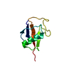
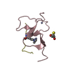
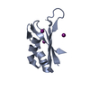
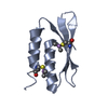
Signal recognition particle alu RNA binding heterodimer, srp9/1 / Signal recognition particle, SRP14 subunit / Signal recognition particle, SRP9/SRP14 subunit / Signal recognition particle 14kD protein / Signal recognition particle alu RNA binding heterodimer, srp9/1 / 2-Layer Sandwich / Alpha Beta Similarity search - Domain/homology
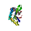
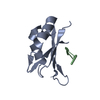
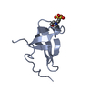
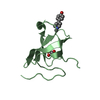
SIGNALRECOGNITIONPARTICLESUBUNITSRP14 / SIGNAL RECOGNITION PARTICLE 14 KDA PROTEIN HOMOLOG / SRP14 FROM SCHIZOSACCHAROMYCES POMBE SIGNAL ...SIGNAL RECOGNITION PARTICLE 14 KDA PROTEIN HOMOLOG / SRP14 FROM SCHIZOSACCHAROMYCES POMBE SIGNAL RECOGNITION PARTICLE
Mass: 10284.420 Da / Num. of mol.: 2 / Fragment: RESIDUES 1-91 Source method: isolated from a genetically manipulated source Details: CYSTEINES 33 AND 78 REACTED WITH CACODYLATE, YIELDING CACODYLATED CYSTEINE ADDUCT (CAS) Source: (gene. exp.) SCHIZOSACCHAROMYCES POMBE (fission yeast) Strain: NCYC 1354 Description: NATIONAL COLLECTION OF YEAST CULTURES, NORWICH, UK Plasmid: PST39 / Production host: ESCHERICHIA COLI (E. coli) / Strain (production host): BL21(AI) / References: UniProt: Q9P372
Resolution: 2.6→5 Å / Num. obs: 5174 / % possible obs: 97.4 % / Observed criterion σ(I): -3 / Redundancy: 5.8 % / Rmerge(I) obs: 0.06 / Net I/σ(I): 16
Reflection shell
Resolution: 2.6→2.7 Å / Redundancy: 6.17 % / Rmerge(I) obs: 0.41 / Mean I/σ(I) obs: 5 / % possible all: 95.5
-
Processing
Software
Name
Version
Classification
REFMAC
5.4.0065
refinement
XDS
datareduction
XSCALE
datascaling
SHELX
phasing
Refinement
Method to determine structure: OTHER Starting model: NONE Resolution: 2.6→32.62 Å / Cor.coef. Fo:Fc: 0.936 / Cor.coef. Fo:Fc free: 0.897 / SU B: 37.967 / SU ML: 0.349 / TLS residual ADP flag: LIKELY RESIDUAL / Cross valid method: THROUGHOUT / ESU R: 1.612 / ESU R Free: 0.367 Stereochemistry target values: MAXIMUM LIKELIHOOD WITH PHASES Details: HYDROGENS HAVE BEEN ADDED IN THE RIDING POSITIONS.
Rfactor
Num. reflection
% reflection
Selection details
Rfree
0.281
236
4.6 %
RANDOM
Rwork
0.238
-
-
-
obs
0.24
4924
97.2 %
-
Solvent computation
Ion probe radii: 0.8 Å / Shrinkage radii: 0.8 Å / VDW probe radii: 1.2 Å / Solvent model: MASK
 Movie
Movie Controller

 Yorodumi
 Open data
 Basic information
 Components
 Keywords
 Function and homology information
 X-RAY DIFFRACTION /
  Authors
 Citation
 Structure visualization
 Downloads & links
 Other downloads
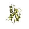


 PDBj
PDBj Assembly
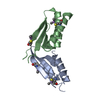


 Mass: 18.015 Da / Num. of mol.: 4 / Source method: isolated from a natural source / Formula: H2O
 Sample preparation
 Processing