 Movie
Movie Controller

[English] 日本語
 Yorodumi
- PDB-1fna: CRYSTAL STRUCTURE OF THE TENTH TYPE III CELL ADHESION MODULE OF H... -
+ Open data

- Basic information
| Entry | Database: PDB / ID: 1fna | ||||||
|---|---|---|---|---|---|---|---|
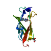
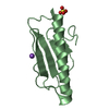
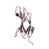
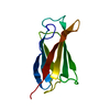
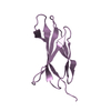
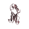
| Title | CRYSTAL STRUCTURE OF THE TENTH TYPE III CELL ADHESION MODULE OF HUMAN FIBRONECTIN | ||||||
 Components Components | FIBRONECTIN CELL-ADHESION MODULE TYPE III-10 | ||||||
 Keywords Keywords | CELL ADHESION PROTEIN | ||||||
| Function / homology |  Function and homology information Function and homology informationnegative regulation of monocyte activation / negative regulation of transforming growth factor beta production / positive regulation of substrate-dependent cell migration, cell attachment to substrate / calcium-independent cell-matrix adhesion / neural crest cell migration involved in autonomic nervous system development / fibrinogen complex / Fibronectin matrix formation / Attachment of bacteria to epithelial cells / enteric nervous system development / integrin activation ...negative regulation of monocyte activation / negative regulation of transforming growth factor beta production / positive regulation of substrate-dependent cell migration, cell attachment to substrate / calcium-independent cell-matrix adhesion / neural crest cell migration involved in autonomic nervous system development / fibrinogen complex / Fibronectin matrix formation / Attachment of bacteria to epithelial cells / enteric nervous system development / integrin activation / ALK mutants bind TKIs / cell-substrate junction assembly / proteoglycan binding / MET activates PTK2 signaling / Molecules associated with elastic fibres / peptidase activator activity / neural crest cell migration / extracellular matrix structural constituent / Syndecan interactions / p130Cas linkage to MAPK signaling for integrins / biological process involved in interaction with symbiont / response to muscle activity / regulation of protein phosphorylation / endoplasmic reticulum-Golgi intermediate compartment / GRB2:SOS provides linkage to MAPK signaling for Integrins / endodermal cell differentiation / Non-integrin membrane-ECM interactions / basement membrane / blood coagulation, fibrin clot formation / ECM proteoglycans / Integrin cell surface interactions / regulation of ERK1 and ERK2 cascade / endothelial cell migration / positive regulation of axon extension / collagen binding / Degradation of the extracellular matrix / Integrin signaling / substrate adhesion-dependent cell spreading / platelet alpha granule lumen / Turbulent (oscillatory, disturbed) flow shear stress activates signaling by PIEZO1 and integrins in endothelial cells / cell-matrix adhesion / acute-phase response / Cell surface interactions at the vascular wall / negative regulation of autophagy / Developmental Lineage of Pancreatic Ductal Cells / integrin-mediated signaling pathway / Post-translational protein phosphorylation / Signaling by high-kinase activity BRAF mutants / MAP2K and MAPK activation / response to wounding / integrin binding / autophagy / Regulation of Insulin-like Growth Factor (IGF) transport and uptake by Insulin-like Growth Factor Binding Proteins (IGFBPs) / positive regulation of fibroblast proliferation / Signaling by RAF1 mutants / Signaling by moderate kinase activity BRAF mutants / Paradoxical activation of RAF signaling by kinase inactive BRAF / Signaling downstream of RAS mutants / Signaling by BRAF and RAF1 fusions / Signaling by ALK fusions and activated point mutants / Platelet degranulation / regulation of cell shape / nervous system development / heparin binding / GPER1 signaling / heart development / extracellular matrix / protease binding / Interleukin-4 and Interleukin-13 signaling / angiogenesis / blood microparticle / positive regulation of phosphatidylinositol 3-kinase/protein kinase B signal transduction / cell adhesion / apical plasma membrane / endoplasmic reticulum lumen / receptor ligand activity / signaling receptor binding / positive regulation of cell population proliferation / positive regulation of gene expression / : / extracellular exosome / extracellular region / identical protein binding / plasma membrane Similarity search - Function | ||||||
| Biological species |  Homo sapiens (human) Homo sapiens (human) | ||||||
| Method |  X-RAY DIFFRACTION / Resolution: 1.8 Å X-RAY DIFFRACTION / Resolution: 1.8 Å | ||||||
 Authors Authors | Dickinson, C.D. / Veerapandian, B. / Ely, K.R. | ||||||
 Citation Citation | Journal: J.Mol.Biol. / Year: 1994 Title: Crystal structure of the tenth type III cell adhesion module of human fibronectin. Authors: Dickinson, C.D. / Veerapandian, B. / Dai, X.P. / Hamlin, R.C. / Xuong, N.H. / Ruoslahti, E. / Ely, K.R. | ||||||
| History |
|
- Structure visualization
| Structure viewer | Molecule: MolmilJmol/JSmol |
|---|
- Downloads & links
-Download
| PDBx/mmCIF format | 1fna.cif.gz | 29.2 KB | Display | PDBx/mmCIF format |
|---|---|---|---|---|
| PDB format | pdb1fna.ent.gz | 20.2 KB | Display | PDB format |
| PDBx/mmJSON format | 1fna.json.gz | Tree view | PDBx/mmJSON format | |
| Others |  Other downloads Other downloads |
-Validation report
| Arichive directory | https://data.pdbj.org/pub/pdb/validation_reports/fn/1fnaftp://data.pdbj.org/pub/pdb/validation_reports/fn/1fna | HTTPS FTP |
|---|
-Related structure data
| Similar structure data |
|---|


-Links
 PDBj
PDBj


- Assembly
| Deposited unit | 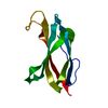
| ||||||||
|---|---|---|---|---|---|---|---|---|---|
| 1 |
| ||||||||
| Unit cell |
|
-Components
| #1: Protein | Mass: 9691.788 Da / Num. of mol.: 1 Source method: isolated from a genetically manipulated source Source: (gene. exp.) Homo sapiens (human) / References: UniProt: P02751 |
|---|
-Experimental details
-Experiment
| Experiment | Method: X-RAY DIFFRACTION |
|---|
- Sample preparation
| Crystal | Density Matthews: 2 Å3/Da / Density % sol: 38.58 % | ||||||||||||
|---|---|---|---|---|---|---|---|---|---|---|---|---|---|
| Crystal grow | *PLUS pH: 8.6 / Method: unknown | ||||||||||||
| Components of the solutions | *PLUS
|
-Data collection
| Reflection | *PLUS Highest resolution: 1.75 Å / Num. obs: 7250 / % possible obs: 96 % / Num. measured all: 34949 / Rmerge(I) obs: 0.072 |
|---|
- Processing
| Software | Name: PROLSQ / Classification: refinement | |||||||||||||||||||||||||||||||||||||||||||||||||||||||||||||||
|---|---|---|---|---|---|---|---|---|---|---|---|---|---|---|---|---|---|---|---|---|---|---|---|---|---|---|---|---|---|---|---|---|---|---|---|---|---|---|---|---|---|---|---|---|---|---|---|---|---|---|---|---|---|---|---|---|---|---|---|---|---|---|---|---|
| Refinement | Resolution: 1.8→10 Å / Rfactor obs: 0.18 | |||||||||||||||||||||||||||||||||||||||||||||||||||||||||||||||
| Refinement step | Cycle: LAST / Resolution: 1.8→10 Å
| |||||||||||||||||||||||||||||||||||||||||||||||||||||||||||||||
| Refine LS restraints |
| |||||||||||||||||||||||||||||||||||||||||||||||||||||||||||||||
| Software | *PLUS Name: PROLSQ / Classification: refinement | |||||||||||||||||||||||||||||||||||||||||||||||||||||||||||||||
| Refinement | *PLUS Highest resolution: 1.8 Å / Lowest resolution: 10 Å / Num. reflection obs: 6996 / Rfactor obs: 0.18 | |||||||||||||||||||||||||||||||||||||||||||||||||||||||||||||||
| Solvent computation | *PLUS | |||||||||||||||||||||||||||||||||||||||||||||||||||||||||||||||
| Displacement parameters | *PLUS Biso mean: 14.6 Å2 | |||||||||||||||||||||||||||||||||||||||||||||||||||||||||||||||
| Refine LS restraints | *PLUS
|
