[English] 日本語
Yorodumi
- PDB-4us7: Sulfur SAD Phased Structure of a Type IV Pilus Protein from Shewa... -

+
Open data


ID or keywords:

Loading...

-
Basic information

Entry
Database: PDB / ID: 4us7
Title
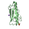
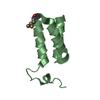
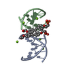
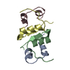
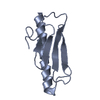
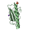
Sulfur SAD Phased Structure of a Type IV Pilus Protein from Shewanella oneidensis
ComponentsPILD PROCESSED PROTEIN
KeywordsCELL ADHESION / TYPE IV PILIN / SULFUR SAD / STRUCTURAL PROTEIN
Function / homology
Function and homology information


type IV pilus / type IV pilus assembly / type IV pilus-dependent motility / membrane
Similarity search - Function
Type IV pilus non-core minor pilin PilE-like / Type IV minor pilin ComP, DNA uptake sequence receptor / Prokaryotic N-terminal methylation site. / Prokaryotic N-terminal methylation motif / Prokaryotic N-terminal methylation site / Pilin-like
Similarity search - Domain/homology
PilD processed protein
Similarity search - Component
Biological speciesSHEWANELLA ONEIDENSIS (bacteria)
MethodX-RAY DIFFRACTION / SYNCHROTRON / SAD / Resolution: 1.96 Å
AuthorsGorgel, M. / Boeggild, A. / Ulstrup, J.J. / Mueller, U. / Weiss, M. / Nissen, P. / Boesen, T.
CitationJournal: Bmc Struct.Biol. / Year: 2015
Title: High-Resolution Structure of a Type Iv Pilin from the Metal- Reducing Bacterium Shewanella Oneidensis.
Authors: Gorgel, M. / Ulstrup, J.J. / Boggild, A. / Jones, N.C. / Hoffmann, S.V. / Nissen, P. / Boesen, T.
History
DepositionJul 3, 2014Deposition site: PDBE / Processing site: PDBE
Revision 1.0Apr 29, 2015Provider: repository / Type: Initial release

-
Structure visualization

Structure viewerMolecule:
MolmilJmol/JSmol

Downloads & links

-
Assembly

Deposited unit
A: PILD PROCESSED PROTEIN
B: PILD PROCESSED PROTEIN
hetero molecules


Theoretical massNumber of molelcules
Total (without water)19,9474
Polymers19,8282
Non-polymers1192
Water2,252125
1
A: PILD PROCESSED PROTEIN


Theoretical massNumber of molelcules
Total (without water)9,9141
Polymers9,9141
Non-polymers00
Water181
TypeNameSymmetry operationNumber
identity operation1_555x,y,z1
MethodPISA
2
B: PILD PROCESSED PROTEIN
hetero molecules


Theoretical massNumber of molelcules
Total (without water)10,0333
Polymers9,9141
Non-polymers1192
Water181
TypeNameSymmetry operationNumber
identity operation1_555x,y,z1
MethodPISA
Unit cell
Length a, b, c (Å)48.830, 96.550, 109.760
Angle α, β, γ (deg.)90.00, 90.00, 90.00
Int Tables number23
Space group name H-MI222

-
Components

#1: Protein PILD PROCESSED PROTEIN / TYPE IV PILUS PROTEIN


Mass: 9913.945 Da / Num. of mol.: 2 / Fragment: UNP RESIDUES 36-123
Source method: isolated from a genetically manipulated source
Details: CYSTEINE BRIDGE BETWEEN CYS69 AND CYS86 IN CHAIN A CYSTEINE BRIDGE BETWEEN CYS69 AND CYS86 IN CHAIN B
Source: (gene. exp.) SHEWANELLA ONEIDENSIS (bacteria) / Strain: MR-1 / Production host: ESCHERICHIA COLI (E. coli) / Strain (production host): BL21(DE3) / Variant (production host): ORIGAMI / References: UniProt: Q8EII5
#2: Chemical ChemComp-SO4 / SULFATE ION


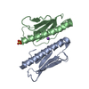
Mass: 96.063 Da / Num. of mol.: 1 / Source method: obtained synthetically / Formula: SO4
#3: Chemical ChemComp-NA / SODIUM ION


Mass: 22.990 Da / Num. of mol.: 1 / Source method: obtained synthetically / Formula: Na
#4: Water ChemComp-HOH / water


Mass: 18.015 Da / Num. of mol.: 125 / Source method: isolated from a natural source / Formula: H2O
Sequence detailsN-TERMINAL G REMAINDER OF TEV CLEAVAGE SITE

-
Experimental details

-
Experiment

ExperimentMethod: X-RAY DIFFRACTION / Number of used crystals: 1

-
Sample preparation

CrystalDensity Matthews: 3.27 Å3/Da / Density % sol: 62.4 % / Description: DATA WERE COLLECTED FOR SULFUR SAD
Crystal growpH: 8 / Details: 28% PEG 8K 100 MM TAPS PH 8.0 150 MM AMS

-
Data collection

DiffractionMean temperature: 100 K
Diffraction sourceSource: SYNCHROTRON / Site: BESSY / Beamline: 14.1 / Wavelength: 1.8
DetectorType: DECTRIS PILATUS 6M / Detector: PIXEL / Date: Jan 24, 2014 / Details: MIRRORS
RadiationMonochromator: SI-111 CRYSTAL / Protocol: SINGLE WAVELENGTH / Monochromatic (M) / Laue (L): M / Scattering type: x-ray
Radiation wavelengthWavelength: 1.8 Å / Relative weight: 1
ReflectionResolution: 1.96→48.8 Å / Num. obs: 34976 / % possible obs: 97.1 % / Observed criterion σ(I): -3 / Redundancy: 17.5 % / Biso Wilson estimate: 27.36 Å2 / Rmerge(I) obs: 0.04 / Net I/σ(I): 51.72
Reflection shellResolution: 1.96→2.01 Å / Redundancy: 13.9 % / Rmerge(I) obs: 0.08 / Mean I/σ(I) obs: 21.15 / % possible all: 94.9

-
Processing

Software
NameVersionClassification
PHENIX(PHENIX.REFINE)refinement
XDSdata reduction
XSCALEdata scaling
PHENIXphasing
RefinementMethod to determine structure: SAD
Starting model: NONE

Resolution: 1.96→48.275 Å / SU ML: 0.18 / σ(F): 1.46 / Phase error: 21.37 / Stereochemistry target values: ML
RfactorNum. reflection% reflection
Rfree0.2201 3473 9.9 %
Rwork0.1794 --
obs0.1835 34971 97.11 %
Solvent computationShrinkage radii: 0.9 Å / VDW probe radii: 1.11 Å / Solvent model: FLAT BULK SOLVENT MODEL
Displacement parametersBiso mean: 34.6 Å2
Refinement stepCycle: LAST / Resolution: 1.96→48.275 Å
ProteinNucleic acidLigandSolventTotal
Num. atoms1400 0 6 125 1531
Refine LS restraints
Refine-IDTypeDev idealNumber
X-RAY DIFFRACTIONf_bond_d0.0061450
X-RAY DIFFRACTIONf_angle_d1.0211969
X-RAY DIFFRACTIONf_dihedral_angle_d11.822525
X-RAY DIFFRACTIONf_chiral_restr0.045201
X-RAY DIFFRACTIONf_plane_restr0.005261
LS refinement shell
Resolution (Å)Rfactor RfreeNum. reflection RfreeRfactor RworkNum. reflection RworkRefine-ID% reflection obs (%)
1.9599-1.98680.23221370.16671220X-RAY DIFFRACTION95
1.9868-2.01520.20321350.16961244X-RAY DIFFRACTION95
2.0152-2.04530.18781360.16691208X-RAY DIFFRACTION95
2.0453-2.07720.24361330.17751241X-RAY DIFFRACTION95
2.0772-2.11130.22711380.1851206X-RAY DIFFRACTION93
2.1113-2.14770.22081390.18751257X-RAY DIFFRACTION96
2.1477-2.18670.26981370.18141213X-RAY DIFFRACTION95
2.1867-2.22880.19411360.17931228X-RAY DIFFRACTION95
2.2288-2.27430.24441320.18431233X-RAY DIFFRACTION96
2.2743-2.32370.29681360.19131244X-RAY DIFFRACTION95
2.3237-2.37780.2231380.19251275X-RAY DIFFRACTION98
2.3778-2.43730.2451390.18481238X-RAY DIFFRACTION95
2.4373-2.50320.27581340.20611271X-RAY DIFFRACTION98
2.5032-2.57680.25741430.20261274X-RAY DIFFRACTION96
2.5768-2.660.25251440.20181290X-RAY DIFFRACTION99
2.66-2.7550.22791380.19051261X-RAY DIFFRACTION97
2.755-2.86530.28151410.20851277X-RAY DIFFRACTION98
2.8653-2.99570.22641410.19281264X-RAY DIFFRACTION99
2.9957-3.15360.231430.20081277X-RAY DIFFRACTION99
3.1536-3.35120.22031390.19131300X-RAY DIFFRACTION99
3.3512-3.60980.19931390.17871274X-RAY DIFFRACTION99
3.6098-3.9730.21241430.16411311X-RAY DIFFRACTION100
3.973-4.54750.19371420.15431297X-RAY DIFFRACTION100
4.5475-5.72790.17691460.15291290X-RAY DIFFRACTION100
5.7279-48.28960.19661440.16981305X-RAY DIFFRACTION100
Refinement TLS params.

Method: refined / Refine-ID: X-RAY DIFFRACTION

IDL112)L122)L132)L222)L232)L332)S11 (Å °)S12 (Å °)S13 (Å °)S21 (Å °)S22 (Å °)S23 (Å °)S31 (Å °)S32 (Å °)S33 (Å °)T112)T122)T132)T222)T232)T332)Origin x (Å)Origin y (Å)Origin z (Å)
15.3556-2.1442-4.04277.43984.73999.17930.18230.01030.00690.1259-0.12550.1132-0.1598-0.64460.07930.1494-0.0108-0.02070.25420.01130.196510.494942.011235.8884
21.6308-0.2498-1.19714.68661.39724.29390.03170.16120.0404-0.10890.0844-0.3743-0.1966-0.151-0.11920.14230.0201-0.01730.1658-0.02870.259118.428445.907933.0302
34.2822-0.6044-0.86772.38340.81827.3489-0.0504-0.19450.58840.93580.21-0.2355-0.228-0.3204-0.1570.41930.0569-0.06790.2207-0.05850.346614.815251.196445.8593
46.3985-0.5544-4.06925.28932.23873.3573-0.4887-0.32310.34111.2360.3145-0.25380.975-0.09170.20060.44830.026-0.07350.2303-0.01560.290315.45239.072245.5571
56.5285-1.1259-2.61725.98152.60488.94150.21050.48140.0812-0.339-0.0820.0196-0.3326-0.1592-0.12190.15250.0297-0.04530.1694-0.00780.22651.84331.915330.1028
63.0278-0.3675-0.59633.22710.3187.01090.14280.412-0.5485-0.13440.0523-0.11390.44390.1226-0.23180.16510.0106-0.05380.2403-0.10190.28237.851224.731129.5191
75.9405-0.49621.45274.80840.75427.65240.0444-0.5181-0.2660.5254-0.16050.2219-0.0396-0.34050.06220.2266-0.03680.01270.19980.00540.2805-2.161425.372138.3835
Refinement TLS group
IDRefine-IDRefine TLS-IDSelection details
1X-RAY DIFFRACTION1CHAIN 'A' AND (RESID 1 THROUGH 27 )
2X-RAY DIFFRACTION2CHAIN 'A' AND (RESID 28 THROUGH 61 )
3X-RAY DIFFRACTION3CHAIN 'A' AND (RESID 62 THROUGH 75 )
4X-RAY DIFFRACTION4CHAIN 'A' AND (RESID 76 THROUGH 89 )
5X-RAY DIFFRACTION5CHAIN 'B' AND (RESID 1 THROUGH 27 )
6X-RAY DIFFRACTION6CHAIN 'B' AND (RESID 28 THROUGH 61 )
7X-RAY DIFFRACTION7CHAIN 'B' AND (RESID 62 THROUGH 89 )

+
About Yorodumi

-
News

-
Feb 9, 2022. New format data for meta-information of EMDB entries

New format data for meta-information of EMDB entries

  • Version 3 of the EMDB header file is now the official format.
  • The previous official version 1.9 will be removed from the archive.

Related info.:EMDB header

External links:wwPDB to switch to version 3 of the EMDB data model

-
Aug 12, 2020. Covid-19 info

Covid-19 info

URL: https://pdbjlc1.pdbj.org/emnavi/covid19.php

New page: Covid-19 featured information page in EM Navigator.

Related info.:Covid-19 info / Mar 5, 2020. Novel coronavirus structure data

+
Mar 5, 2020. Novel coronavirus structure data

Novel coronavirus structure data

Related info.:Yorodumi Speices / Aug 12, 2020. Covid-19 info

External links:COVID-19 featured content - PDBj / Molecule of the Month (242):Coronavirus Proteases

+
Jan 31, 2019. EMDB accession codes are about to change! (news from PDBe EMDB page)

EMDB accession codes are about to change! (news from PDBe EMDB page)

  • The allocation of 4 digits for EMDB accession codes will soon come to an end. Whilst these codes will remain in use, new EMDB accession codes will include an additional digit and will expand incrementally as the available range of codes is exhausted. The current 4-digit format prefixed with “EMD-” (i.e. EMD-XXXX) will advance to a 5-digit format (i.e. EMD-XXXXX), and so on. It is currently estimated that the 4-digit codes will be depleted around Spring 2019, at which point the 5-digit format will come into force.
  • The EM Navigator/Yorodumi systems omit the EMD- prefix.

Related info.:Q: What is EMD? / ID/Accession-code notation in Yorodumi/EM Navigator

External links:EMDB Accession Codes are Changing Soon! / Contact to PDBj

+
Jul 12, 2017. Major update of PDB

Major update of PDB

  • wwPDB released updated PDB data conforming to the new PDBx/mmCIF dictionary.
  • This is a major update changing the version number from 4 to 5, and with Remediation, in which all the entries are updated.
  • In this update, many items about electron microscopy experimental information are reorganized (e.g. em_software).
  • Now, EM Navigator and Yorodumi are based on the updated data.

External links:wwPDB Remediation / Enriched Model Files Conforming to OneDep Data Standards Now Available in the PDB FTP Archive

-
Yorodumi

Thousand views of thousand structures

  • Yorodumi is a browser for structure data from EMDB, PDB, SASBDB, etc.
  • This page is also the successor to EM Navigator detail page, and also detail information page/front-end page for Omokage search.
  • The word "yorodu" (or yorozu) is an old Japanese word meaning "ten thousand". "mi" (miru) is to see.

Related info.:EMDB / PDB / SASBDB / Comparison of 3 databanks / Yorodumi Search / Aug 31, 2016. New EM Navigator & Yorodumi / Yorodumi Papers / Jmol/JSmol / Function and homology information / Changes in new EM Navigator and Yorodumi

Read more