 Movie
Movie Controller


+ Open data

- Basic information
| Entry | Database: PDB / ID: 5mix | ||||||
|---|---|---|---|---|---|---|---|
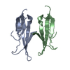
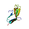
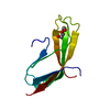
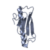
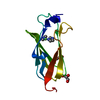
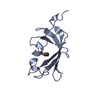
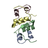
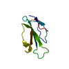
| Title | Extracellular domain of human CD83 - trigonal crystal form | ||||||
 Components Components | CD83 antigen | ||||||
 Keywords Keywords | IMMUNE SYSTEM / Dendritic cell / receptor / immunoglobulin | ||||||
| Function / homology |  Function and homology information Function and homology informationpositive regulation of CD4-positive, alpha-beta T cell differentiation / negative regulation of interleukin-4 production / CD4-positive, alpha-beta T cell differentiation / humoral immune response / positive regulation of interleukin-10 production / positive regulation of interleukin-2 production / defense response / external side of plasma membrane / signal transduction / plasma membrane Similarity search - Function | ||||||
| Biological species |  Homo sapiens (human) Homo sapiens (human) | ||||||
| Method |  X-RAY DIFFRACTION / SYNCHROTRON / Resolution: 1.701 Å X-RAY DIFFRACTION / SYNCHROTRON / Resolution: 1.701 Å | ||||||
 Authors Authors | Klingl, S. / Egerer-Sieber, C. / Schmid, B. / Muller, Y.A. | ||||||
| Funding support |  Germany, 1items Germany, 1items
| ||||||
 Citation Citation | Journal: J. Mol. Biol. / Year: 2017 Title: Crystal Structure of the Extracellular Domain of the Human Dendritic Cell Surface Marker CD83. Authors: Heilingloh, C.S. / Klingl, S. / Egerer-Sieber, C. / Schmid, B. / Weiler, S. / Muhl-Zurbes, P. / Hofmann, J. / Stump, J.D. / Sticht, H. / Kummer, M. / Steinkasserer, A. / Muller, Y.A. | ||||||
| History |
|
- Structure visualization
| Structure viewer | Molecule: MolmilJmol/JSmol |
|---|
- Downloads & links
-Download
| PDBx/mmCIF format | 5mix.cif.gz | 49.9 KB | Display | PDBx/mmCIF format |
|---|---|---|---|---|
| PDB format | pdb5mix.ent.gz | 34.6 KB | Display | PDB format |
| PDBx/mmJSON format | 5mix.json.gz | Tree view | PDBx/mmJSON format | |
| Others |  Other downloads Other downloads |
-Validation report
| Arichive directory | https://data.pdbj.org/pub/pdb/validation_reports/mi/5mixftp://data.pdbj.org/pub/pdb/validation_reports/mi/5mix | HTTPS FTP |
|---|
-Related structure data


-Links
 PDBj
PDBj- Assembly
| Deposited unit | 
| ||||||||
|---|---|---|---|---|---|---|---|---|---|
| 1 |
| ||||||||
| Unit cell |
|
-Components
| #1: Protein | Mass: 12766.979 Da / Num. of mol.: 1 / Mutation: C27S, C100S, C129S Source method: isolated from a genetically manipulated source Details: The first four residues (GSPG) are non-native residues of the linker which remains after the GST-tag was cleaved off. The first three and last four residues as well as the central region ...Details: The first four residues (GSPG) are non-native residues of the linker which remains after the GST-tag was cleaved off. The first three and last four residues as well as the central region were not visible in the electron density maps. Source: (gene. exp.) Homo sapiens (human) / Cell: Dendritic cells / Gene: CD83 / Plasmid: pGEX-2T / Production host:  |
|---|---|
| #2: Water | ChemComp-HOH /  Mass: 18.015 Da / Num. of mol.: 31 / Source method: isolated from a natural source / Formula: H2O Mass: 18.015 Da / Num. of mol.: 31 / Source method: isolated from a natural source / Formula: H2O |
| Has protein modification | Y |
-Experimental details
-Experiment
| Experiment | Method: X-RAY DIFFRACTION / Number of used crystals: 1 |
|---|
- Sample preparation
| Crystal | Density Matthews: 1.79 Å3/Da / Density % sol: 31.45 % |
|---|---|
| Crystal grow | Temperature: 293 K / Method: vapor diffusion, sitting drop Details: 0.4 microliter protein (39 mg/ml in 50 mM Tris-HCl (pH 8.0) buffer) were mixed with 0.2 microliter reservoir solution (1 M ammonium sulfate, 0.1 M HEPES (pH 7.0), 0.5% w/v polyethylene ...Details: 0.4 microliter protein (39 mg/ml in 50 mM Tris-HCl (pH 8.0) buffer) were mixed with 0.2 microliter reservoir solution (1 M ammonium sulfate, 0.1 M HEPES (pH 7.0), 0.5% w/v polyethylene glycol (PEG) 8000) and equilibrated against 70 microliter of reservoir solution |
-Data collection
| Diffraction | Mean temperature: 100 K |
|---|---|
| Diffraction source | Source: SYNCHROTRON / Site: BESSY / Beamline: 14.1 / Wavelength: 0.918409 Å |
| Detector | Type: DECTRIS PILATUS 6M / Detector: PIXEL / Date: May 30, 2015 |
| Radiation | Protocol: SINGLE WAVELENGTH / Monochromatic (M) / Laue (L): M / Scattering type: x-ray |
| Radiation wavelength | Wavelength: 0.918409 Å / Relative weight: 1 |
| Reflection | Resolution: 1.7→40.06 Å / Num. obs: 10348 / % possible obs: 99.9 % / Redundancy: 9.9 % / Biso Wilson estimate: 38.5 Å2 / Rmerge(I) obs: 0.046 / Net I/σ(I): 23.8 |
| Reflection shell | Resolution: 1.7→1.8 Å / Redundancy: 9.9 % / Rmerge(I) obs: 0.851 / Mean I/σ(I) obs: 2.6 / % possible all: 99.9 |
- Processing
| Software |
| |||||||||||||||||||||||||||||||||||||||||||||||||||||||||||||||||||||||||||||||||||||||||||||||||||||||||||||||||||||||||||||||||||||||||||||||||||||||||||||||||||||||||||||||||||||||||||||||||||||||||||||||||||||||||||||||||||||||||||||||||||||||||||||||||||||||||||||||||||
|---|---|---|---|---|---|---|---|---|---|---|---|---|---|---|---|---|---|---|---|---|---|---|---|---|---|---|---|---|---|---|---|---|---|---|---|---|---|---|---|---|---|---|---|---|---|---|---|---|---|---|---|---|---|---|---|---|---|---|---|---|---|---|---|---|---|---|---|---|---|---|---|---|---|---|---|---|---|---|---|---|---|---|---|---|---|---|---|---|---|---|---|---|---|---|---|---|---|---|---|---|---|---|---|---|---|---|---|---|---|---|---|---|---|---|---|---|---|---|---|---|---|---|---|---|---|---|---|---|---|---|---|---|---|---|---|---|---|---|---|---|---|---|---|---|---|---|---|---|---|---|---|---|---|---|---|---|---|---|---|---|---|---|---|---|---|---|---|---|---|---|---|---|---|---|---|---|---|---|---|---|---|---|---|---|---|---|---|---|---|---|---|---|---|---|---|---|---|---|---|---|---|---|---|---|---|---|---|---|---|---|---|---|---|---|---|---|---|---|---|---|---|---|---|---|---|---|---|---|---|---|---|---|---|---|---|---|---|---|---|---|---|---|---|---|---|---|---|---|---|---|---|---|---|---|---|---|---|---|---|---|---|---|---|---|---|---|---|---|---|---|---|---|---|---|---|---|
| Refinement | Resolution: 1.701→40.06 Å / SU ML: 0.22 / Cross valid method: FREE R-VALUE / σ(F): 1.38 / Phase error: 25.13
| |||||||||||||||||||||||||||||||||||||||||||||||||||||||||||||||||||||||||||||||||||||||||||||||||||||||||||||||||||||||||||||||||||||||||||||||||||||||||||||||||||||||||||||||||||||||||||||||||||||||||||||||||||||||||||||||||||||||||||||||||||||||||||||||||||||||||||||||||||
| Solvent computation | Shrinkage radii: 0.9 Å / VDW probe radii: 1.11 Å | |||||||||||||||||||||||||||||||||||||||||||||||||||||||||||||||||||||||||||||||||||||||||||||||||||||||||||||||||||||||||||||||||||||||||||||||||||||||||||||||||||||||||||||||||||||||||||||||||||||||||||||||||||||||||||||||||||||||||||||||||||||||||||||||||||||||||||||||||||
| Displacement parameters | Biso mean: 44.6 Å2 | |||||||||||||||||||||||||||||||||||||||||||||||||||||||||||||||||||||||||||||||||||||||||||||||||||||||||||||||||||||||||||||||||||||||||||||||||||||||||||||||||||||||||||||||||||||||||||||||||||||||||||||||||||||||||||||||||||||||||||||||||||||||||||||||||||||||||||||||||||
| Refinement step | Cycle: LAST / Resolution: 1.701→40.06 Å
| |||||||||||||||||||||||||||||||||||||||||||||||||||||||||||||||||||||||||||||||||||||||||||||||||||||||||||||||||||||||||||||||||||||||||||||||||||||||||||||||||||||||||||||||||||||||||||||||||||||||||||||||||||||||||||||||||||||||||||||||||||||||||||||||||||||||||||||||||||
| Refine LS restraints |
| |||||||||||||||||||||||||||||||||||||||||||||||||||||||||||||||||||||||||||||||||||||||||||||||||||||||||||||||||||||||||||||||||||||||||||||||||||||||||||||||||||||||||||||||||||||||||||||||||||||||||||||||||||||||||||||||||||||||||||||||||||||||||||||||||||||||||||||||||||
| LS refinement shell |
| |||||||||||||||||||||||||||||||||||||||||||||||||||||||||||||||||||||||||||||||||||||||||||||||||||||||||||||||||||||||||||||||||||||||||||||||||||||||||||||||||||||||||||||||||||||||||||||||||||||||||||||||||||||||||||||||||||||||||||||||||||||||||||||||||||||||||||||||||||
| Refinement TLS params. | Method: refined / Refine-ID: X-RAY DIFFRACTION
| |||||||||||||||||||||||||||||||||||||||||||||||||||||||||||||||||||||||||||||||||||||||||||||||||||||||||||||||||||||||||||||||||||||||||||||||||||||||||||||||||||||||||||||||||||||||||||||||||||||||||||||||||||||||||||||||||||||||||||||||||||||||||||||||||||||||||||||||||||
| Refinement TLS group |
|
