- PDB-5mj0: Extracellular domain of human CD83 - cubic crystal form -
+
Open data
ID or keywords:
Loading...
-
Basic information
Entry
Database: PDB / ID: 5mj0
Title
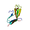
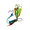
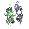
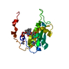
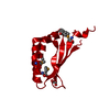
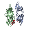
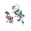
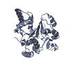
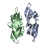
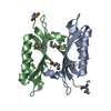
Extracellular domain of human CD83 - cubic crystal form
Components
CD83 antigen
Keywords
IMMUNE SYSTEM / Dendritic cell / receptor / immunoglobulin
Function / homology
Function and homology information
positive regulation of CD4-positive, alpha-beta T cell differentiation / negative regulation of interleukin-4 production / CD4-positive, alpha-beta T cell differentiation / humoral immune response / positive regulation of interleukin-10 production / positive regulation of interleukin-2 production / defense response / external side of plasma membrane / signal transduction / plasma membrane Similarity search - Function
CD83antigen / hCD83 / B-cell activation protein / Cell surface protein HB15
Mass: 12766.979 Da / Num. of mol.: 2 / Mutation: C27S, C100S, C129S Source method: isolated from a genetically manipulated source Details: The first four residues (GSPG) are non-native residues of the linker which remains after the GST-tag was cleaved off. The first and last two residues as well as the central region were not ...Details: The first four residues (GSPG) are non-native residues of the linker which remains after the GST-tag was cleaved off. The first and last two residues as well as the central region were not visible in the electron density maps. Source: (gene. exp.) Homo sapiens (human) / Cell: Dendritic cells / Gene: CD83 / Plasmid: pGEX-2T / Production host: Escherichia coli 'BL21-Gold(DE3)pLysS AG' / References: UniProt: Q01151
Has protein modification
Y
-
Experimental details
-
Experiment
Experiment
Method: X-RAY DIFFRACTION / Number of used crystals: 1
-
Sample preparation
Crystal
Density Matthews: 2.73 Å3/Da / Density % sol: 54.94 %
Crystal grow
Temperature: 293 K / Method: vapor diffusion, sitting drop Details: 0.2 microliter protein (21 mg/ml in 25 mM Tris-HCl (pH 7.2) buffer) were mixed with 0.2 microliter reservoir solution (0.2 M L-proline, 0.1 M HEPES (pH 7.5), 24% w/v PEG 1500) and ...Details: 0.2 microliter protein (21 mg/ml in 25 mM Tris-HCl (pH 7.2) buffer) were mixed with 0.2 microliter reservoir solution (0.2 M L-proline, 0.1 M HEPES (pH 7.5), 24% w/v PEG 1500) and equilibrated against 70 microliter of reservoir solution Crystals appeared after ~ 10 months
In the structure databanks used in Yorodumi, some data are registered as the other names, "COVID-19 virus" and "2019-nCoV". Here are the details of the virus and the list of structure data.
Jan 31, 2019. EMDB accession codes are about to change! (news from PDBe EMDB page)
EMDB accession codes are about to change! (news from PDBe EMDB page)
The allocation of 4 digits for EMDB accession codes will soon come to an end. Whilst these codes will remain in use, new EMDB accession codes will include an additional digit and will expand incrementally as the available range of codes is exhausted. The current 4-digit format prefixed with “EMD-” (i.e. EMD-XXXX) will advance to a 5-digit format (i.e. EMD-XXXXX), and so on. It is currently estimated that the 4-digit codes will be depleted around Spring 2019, at which point the 5-digit format will come into force.
The EM Navigator/Yorodumi systems omit the EMD- prefix.
Related info.:Q: What is EMD? / ID/Accession-code notation in Yorodumi/EM Navigator
Yorodumi is a browser for structure data from EMDB, PDB, SASBDB, etc.
This page is also the successor to EM Navigator detail page, and also detail information page/front-end page for Omokage search.
The word "yorodu" (or yorozu) is an old Japanese word meaning "ten thousand". "mi" (miru) is to see.
Related info.:EMDB / PDB / SASBDB / Comparison of 3 databanks / Yorodumi Search / Aug 31, 2016. New EM Navigator & Yorodumi / Yorodumi Papers / Jmol/JSmol / Function and homology information / Changes in new EM Navigator and Yorodumi
 Movie
Movie Controller


 Open data
 Basic information
 Components
 Keywords
 Function and homology information
 Homo sapiens (human)
 X-RAY DIFFRACTION /
  Authors
 Germany, 1items
  Citation
 Structure visualization
 Downloads & links
 Other downloads
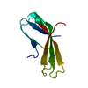
 PDBj
PDBj Assembly
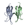
 Sample preparation
 Processing