 Movie
Movie Controller

[English] 日本語
 Yorodumi
- PDB-2pys: Crystal Structure of a Five Site Mutated Cyanovirin-N with a Mann... -
+ Open data

- Basic information
| Entry | Database: PDB / ID: 2pys | |||||||||
|---|---|---|---|---|---|---|---|---|---|---|
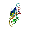
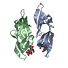
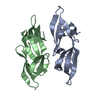
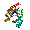
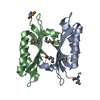
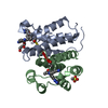
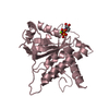
| Title | Crystal Structure of a Five Site Mutated Cyanovirin-N with a Mannose Dimer Bound at 1.8 A Resolution | |||||||||
 Components Components | Cyanovirin-N | |||||||||
 Keywords Keywords | SUGAR BINDING PROTEIN / CYANOVIRIN-N / ANTI HIV | |||||||||
| Function / homology |  Function and homology information Function and homology informationoligosaccharide binding / regulation of defense response to virus / carbohydrate binding Similarity search - Function | |||||||||
| Biological species |  Nostoc ellipsosporum (bacteria) Nostoc ellipsosporum (bacteria) | |||||||||
| Method |  X-RAY DIFFRACTION / MOLECULAR REPLACEMENT / Resolution: 1.8 Å X-RAY DIFFRACTION / MOLECULAR REPLACEMENT / Resolution: 1.8 Å | |||||||||
 Authors Authors | Fromme, R. / Katilene, Z. / Fromme, P. / Ghirlanda, G. | |||||||||
 Citation Citation | Journal: Biochemistry / Year: 2007 Title: A Monovalent Mutant of Cyanovirin-N Provides Insight into the Role of Multiple Interactions with gp120 for Antiviral Activity. Authors: Fromme, R. / Katiliene, Z. / Giomarelli, B. / Bogani, F. / Mahon, J.M. / Mori, T. / Fromme, P. / Ghirlanda, G. | |||||||||
| History |
|
- Structure visualization
| Structure viewer | Molecule: MolmilJmol/JSmol |
|---|
- Downloads & links
-Download
| PDBx/mmCIF format | 2pys.cif.gz | 97.3 KB | Display | PDBx/mmCIF format |
|---|---|---|---|---|
| PDB format | pdb2pys.ent.gz | 73.8 KB | Display | PDB format |
| PDBx/mmJSON format | 2pys.json.gz | Tree view | PDBx/mmJSON format | |
| Others |  Other downloads Other downloads |
-Validation report
| Arichive directory | https://data.pdbj.org/pub/pdb/validation_reports/py/2pysftp://data.pdbj.org/pub/pdb/validation_reports/py/2pys | HTTPS FTP |
|---|
-Related structure data
| Related structure data |  2z21C  1lomS  2pyv S: Starting model for refinement C: citing same article ( |
|---|---|
| Similar structure data |


-Links
 PDBj
PDBj

- Assembly
| Deposited unit | 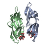
| ||||||||
|---|---|---|---|---|---|---|---|---|---|
| 1 |
| ||||||||
| Unit cell |
|
-Components
| #1: Protein | Mass: 11949.095 Da / Num. of mol.: 2 / Mutation: K3N, T7A, E23I, P51G, N93A Source method: isolated from a genetically manipulated source Source: (gene. exp.) Nostoc ellipsosporum (bacteria) / Gene: CYANOVIRN-N / Production host: #2: Polysaccharide |   Source method: isolated from a genetically manipulated source Details: oligosaccharide / References: 2alpha-alpha-mannobiose #3: Water | ChemComp-HOH / |  Mass: 18.015 Da / Num. of mol.: 184 / Source method: isolated from a natural source / Formula: H2O Mass: 18.015 Da / Num. of mol.: 184 / Source method: isolated from a natural source / Formula: H2OHas protein modification | Y | |
|---|
-Experimental details
-Experiment
| Experiment | Method: X-RAY DIFFRACTION / Number of used crystals: 1 |
|---|
- Sample preparation
| Crystal | Density Matthews: 2.17 Å3/Da / Density % sol: 43.27 % |
|---|---|
| Crystal grow | Temperature: 298 K / Method: vapor diffusion, hanging drop / pH: 6 Details: 30% PEG 8000, 100 MM MGSO4, 2 MM MAN2, pH 6.00, VAPOR DIFFUSION, HANGING DROP, temperature 298K |
-Data collection
| Diffraction | Mean temperature: 100 K |
|---|---|
| Diffraction source | Source: ROTATING ANODE / Type: RIGAKU / Wavelength: 1.5418 |
| Detector | Type: RIGAKU RAXIS IV / Detector: IMAGE PLATE / Date: Nov 9, 2005 / Details: MIRRORS |
| Radiation | Protocol: SINGLE WAVELENGTH / Monochromatic (M) / Laue (L): M / Scattering type: x-ray |
| Radiation wavelength | Wavelength: 1.5418 Å / Relative weight: 1 |
| Reflection | Resolution: 1.8→55.22 Å / Num. obs: 19361 / % possible obs: 87.7 % / Observed criterion σ(I): 4 / Redundancy: 1.9 % / Rmerge(I) obs: 0.094 / Net I/σ(I): 8.13 |
| Reflection shell | Resolution: 1.8→1.85 Å / Redundancy: 1.4 % / Rmerge(I) obs: 0.244 / Mean I/σ(I) obs: 2.92 / % possible all: 82.33 |
- Processing
| Software |
| ||||||||||||||||||||||||||||||||||||||||||||||||||||||||||||||||||||||||||||||||||||||||||||||||||||||||||||||||||||||||||||||||||||||||||||
|---|---|---|---|---|---|---|---|---|---|---|---|---|---|---|---|---|---|---|---|---|---|---|---|---|---|---|---|---|---|---|---|---|---|---|---|---|---|---|---|---|---|---|---|---|---|---|---|---|---|---|---|---|---|---|---|---|---|---|---|---|---|---|---|---|---|---|---|---|---|---|---|---|---|---|---|---|---|---|---|---|---|---|---|---|---|---|---|---|---|---|---|---|---|---|---|---|---|---|---|---|---|---|---|---|---|---|---|---|---|---|---|---|---|---|---|---|---|---|---|---|---|---|---|---|---|---|---|---|---|---|---|---|---|---|---|---|---|---|---|---|---|
| Refinement | Method to determine structure: MOLECULAR REPLACEMENT Starting model: pdb entry 1LOM Resolution: 1.8→9.99 Å / Cor.coef. Fo:Fc: 0.963 / Cor.coef. Fo:Fc free: 0.94 / SU B: 6.604 / SU ML: 0.09 / Cross valid method: THROUGHOUT / ESU R: 0.374 / ESU R Free: 0.129 Stereochemistry target values: MAXIMUM LIKELIHOOD WITH PHASES Details: HYDROGENS HAVE BEEN ADDED IN THE RIDING POSITIONS
| ||||||||||||||||||||||||||||||||||||||||||||||||||||||||||||||||||||||||||||||||||||||||||||||||||||||||||||||||||||||||||||||||||||||||||||
| Solvent computation | Ion probe radii: 0.8 Å / Shrinkage radii: 0.8 Å / VDW probe radii: 1.4 Å / Solvent model: BABINET MODEL WITH MASK | ||||||||||||||||||||||||||||||||||||||||||||||||||||||||||||||||||||||||||||||||||||||||||||||||||||||||||||||||||||||||||||||||||||||||||||
| Displacement parameters | Biso mean: 22.49 Å2
| ||||||||||||||||||||||||||||||||||||||||||||||||||||||||||||||||||||||||||||||||||||||||||||||||||||||||||||||||||||||||||||||||||||||||||||
| Refinement step | Cycle: LAST / Resolution: 1.8→9.99 Å
| ||||||||||||||||||||||||||||||||||||||||||||||||||||||||||||||||||||||||||||||||||||||||||||||||||||||||||||||||||||||||||||||||||||||||||||
| Refine LS restraints |
| ||||||||||||||||||||||||||||||||||||||||||||||||||||||||||||||||||||||||||||||||||||||||||||||||||||||||||||||||||||||||||||||||||||||||||||
| LS refinement shell | Resolution: 1.8→1.85 Å / Total num. of bins used: 20
|
