 Movie
Movie Controller

[English] 日本語
 Yorodumi
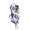
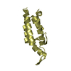
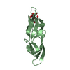
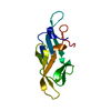
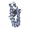
- PDB-2ezm: SOLUTION NMR STRUCTURE OF CYANOVIRIN-N, RESTRAINED REGULARIZED ME... -
+ Open data

- Basic information
| Entry | Database: PDB / ID: 2ezm | ||||||
|---|---|---|---|---|---|---|---|
| Title | SOLUTION NMR STRUCTURE OF CYANOVIRIN-N, RESTRAINED REGULARIZED MEAN COORDINATES | ||||||
 Components Components | CYANOVIRIN-N | ||||||
 Keywords Keywords | HIV-INACTIVATING PROTEIN | ||||||
| Function / homology |  Function and homology information Function and homology informationoligosaccharide binding / regulation of defense response to virus / carbohydrate binding Similarity search - Function | ||||||
| Biological species |  Nostoc ellipsosporum (bacteria) Nostoc ellipsosporum (bacteria) | ||||||
| Method | SOLUTION NMR / simulated annealing | ||||||
 Authors Authors | Bewley, C.A. / Gronenborn, A.M. / Clore, G.M. | ||||||
 Citation Citation | Journal: Nat.Struct.Biol. / Year: 1998 Title: Solution structure of cyanovirin-N, a potent HIV-inactivating protein. Authors: Bewley, C.A. / Gustafson, K.R. / Boyd, M.R. / Covell, D.G. / Bax, A. / Clore, G.M. / Gronenborn, A.M. | ||||||
| History |
|
- Structure visualization
| Structure viewer | Molecule: MolmilJmol/JSmol |
|---|
- Downloads & links
-Download
| PDBx/mmCIF format | 2ezm.cif.gz | 43.9 KB | Display | PDBx/mmCIF format |
|---|---|---|---|---|
| PDB format | pdb2ezm.ent.gz | 31.3 KB | Display | PDB format |
| PDBx/mmJSON format | 2ezm.json.gz | Tree view | PDBx/mmJSON format | |
| Others |  Other downloads Other downloads |
-Validation report
| Arichive directory | https://data.pdbj.org/pub/pdb/validation_reports/ez/2ezmftp://data.pdbj.org/pub/pdb/validation_reports/ez/2ezm | HTTPS FTP |
|---|
-Related structure data


-Links
 PDBj
PDBj- Assembly
| Deposited unit | 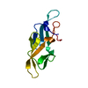
| |||||||||
|---|---|---|---|---|---|---|---|---|---|---|
| 1 |
| |||||||||
| NMR ensembles |
|
-Components
| #1: Protein | Mass: 11022.090 Da / Num. of mol.: 1 / Source method: isolated from a natural source / Source: (natural) Nostoc ellipsosporum (bacteria) / References: UniProt: P81180 |
|---|---|
| Has protein modification | Y |
-Experimental details
-Experiment
| Experiment | Method: SOLUTION NMR | ||||||||||||||||||||||||||||||||||||||||||||||||||||||||||||||||||||||||
|---|---|---|---|---|---|---|---|---|---|---|---|---|---|---|---|---|---|---|---|---|---|---|---|---|---|---|---|---|---|---|---|---|---|---|---|---|---|---|---|---|---|---|---|---|---|---|---|---|---|---|---|---|---|---|---|---|---|---|---|---|---|---|---|---|---|---|---|---|---|---|---|---|---|
| NMR experiment |
| ||||||||||||||||||||||||||||||||||||||||||||||||||||||||||||||||||||||||
| NMR details | Text: THE 3D STRUCTURE OF CYANOVIRIN SOLVED BY MULTI-DIMENSIONAL HETERONUCLEAR NMR AND IS BASED ON 2597 EXPERIMENTAL NMR RESTRAINTS: 419 SEQUENTIAL (|I- J|=1), 170 MEDIUM RANGE (1 < |I-J| 5) ...Text: THE 3D STRUCTURE OF CYANOVIRIN SOLVED BY MULTI-DIMENSIONAL HETERONUCLEAR NMR AND IS BASED ON 2597 EXPERIMENTAL NMR RESTRAINTS: 419 SEQUENTIAL (|I- J|=1), 170 MEDIUM RANGE (1 < |I-J| <=5) AND 554 LONG RANGE (|I-J| >5) INTERRESIDUES AND 19 INTRARESIDUE APPROXIMATE INTERPROTON DISTANCE RESTRAINTS; 109 DISTANCE RESTRAINTS FOR 55 H-BONDS; 339 TORSION ANGLE RESTRAINTS (100 PHI, 98 PSI, 76 CHI1, 48 CHI2, 15 CHI3, 2 CHI4); 82 THREE-BOND HN-HA COUPLING CONSTANT RESTRAINTS; 157 (82 CALPHA AND 75 CBETA) 13C SHIFT RESTRAINTS; 362 1H SHIFT RESTRAINTS; AND 386 DIPOLAR COUPLING RESTRAINTS (82 N-H, 76 C-H, 43 CA-C', 65 N-C' 62 HNC', 58 SIDE-CHAIN C-H). THE STRUCTURES WERE CALCULATED USING THE SIMULATED ANNEALING PROTOCOL OF NILGES ET AL. (1988) FEBS LETT. 229, 129-136 USING THE PROGRAM CNS (BRUNGER ET AL. ACTA CRYST SERIES D IN PRESS) MODIFIED TO INCORPORATE COUPLING CONSTANT (GARRETT ET AL. (1984) J. MAGN. RESON. SERIES B 104, 99-103), CARBON CHEMICAL SHIFT (KUSZEWSKI ET AL. (1995) J. MAGN. RESON. SERIES B 106, 92-96), 1H CHEMICAL SHIFT (KUSZEWSKI ET AL. (1995) J. MAGN. RESON. SERIES B 107, 293-297; KUSZEWSKI ET AL. (1996) J. MAGN. RESON. SERIES B 112, 79-81), AND DIPOLAR COUPLING (CLORE ET AL. (1998) J. MAGN. RESON. 131, 159-162) RESTRAINTS, AND A CONFORMATIONAL DATABASE POTENTIAL (KUSZEWSKI ET AL. (1996) PROTEIN SCI. 5, 1067-1080; KUSZEWSKI ET AL. (1997) J. MAGN. RESON 125, 171-177). |
- Sample preparation
| Sample conditions | pH: 6.1 / Temperature: 300 K |
|---|---|
| Crystal grow | *PLUS Method: other / Details: NMR |
-NMR measurement
| NMR spectrometer |
|
|---|
- Processing
| Software |
| |||||||||
|---|---|---|---|---|---|---|---|---|---|---|
| NMR software |
| |||||||||
| Refinement | Method: simulated annealing / Software ordinal: 1 Details: AVE.RMS DIFF. TO MEAN FOR ALL NON-H-ATOMS (RESIDUES 1:101)= 0.433636 AVE.RMS DIFF. TO MEAN FOR BACKBONE ATOMS (N, CA, C', O) (RESIDUES 1:101)= 0.139826 RMS DEVIATIONS FOR BONDS, ANGLES, ...Details: AVE.RMS DIFF. TO MEAN FOR ALL NON-H-ATOMS (RESIDUES 1:101)= 0.433636 AVE.RMS DIFF. TO MEAN FOR BACKBONE ATOMS (N, CA, C', O) (RESIDUES 1:101)= 0.139826 RMS DEVIATIONS FOR BONDS, ANGLES, IMPROPERS, CDIH, NOE, COUP 5.067337E-03, 0.712983, 0.667194, 0.157308, 1.428009E-02, 0.608909 C13CA AND CB SHIFTS RMS : 0.852581, 1.15742 JCOUP STATS: NON-GLY RESIDUES GLY RMS-D: 0.608909 1.46813 BACKBONE DIPOLAR COUPLINGS NH CH CACO NCO HNCO RMS : 0.466712 1.15058 1.29414 0.572019 1.2653 SIDECHAIN DIPOLAR COUPLINGS CH CH3S CH3D ARO RMS DIPO_SIDE: 1.6875 0.796796 0.531907 0.160016 RMS FOR 1H SHIFTS: ALL ALPHA ALPHA_GLY METHYL(S) METHYL(D) OTHER(S) OTHER(D) RMS PROT: 0.263524 0.243015 0.23853 0.115892 0.148148 0.267304 0.300122 IN THE RESTRAINED REGULARIZED MEAN COORDINATES (2EZM) THE LAST COLUMN REPRESENTS THE AVERAGE RMS DIFFERENCE BETWEEN THE INDIVIDUAL SIMULATED ANNEALING STRUCTURES AND THE MEAN COORDINATE POSITIONS. THE LAST COLUMN IN THE INDIVIDUAL SA STRUCTURES (2EZN) HAS NO MEANING. BEST FITTING TO GENERATE THE AVERAGE STRUCTURE IS WITH RESPECT TO RESIDUES 1-101. NOTE THE OCCUPANCY FIELD HAS NO MEANING. | |||||||||
| NMR ensemble | Conformer selection criteria: REGULARIZED MEAN STRUCTURE / Conformers submitted total number: 1 |
