 Movie
Movie Controller


+ Open data

- Basic information
| Entry | Database: PDB / ID: 1l5e | ||||||
|---|---|---|---|---|---|---|---|
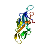
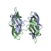
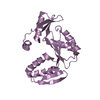
| Title | The domain-swapped dimer of CV-N in solution | ||||||
 Components Components | Cyanovirin-N | ||||||
 Keywords Keywords | ANTIVIRAL PROTEIN / 3D domain-swapping / cyanovirin-N / protein folding | ||||||
| Function / homology |  Function and homology information Function and homology informationoligosaccharide binding / regulation of defense response to virus / carbohydrate binding Similarity search - Function | ||||||
| Biological species |  Nostoc ellipsosporum (bacteria) Nostoc ellipsosporum (bacteria) | ||||||
| Method | SOLUTION NMR / Determination of the domain orientation for the solution structure of the dimer was carried out using a procedure analogous to the one described for determining the relative domain orientation in a two-domain protein fragment of a lectin. | ||||||
 Authors Authors | Barrientos, L.G. / Louis, J.M. / Botos, I. / Mori, T. / Han, Z. / O'Keefe, B.R. / Boyd, M.R. / Wlodawer, A. / Gronenborn, A.M. | ||||||
 Citation Citation | Journal: Structure / Year: 2002 Title: The domain-swapped dimer of cyanovirin-N is in a metastable folded state: reconciliation of X-ray and NMR structures. Authors: Barrientos, L.G. / Louis, J.M. / Botos, I. / Mori, T. / Han, Z. / O'Keefe, B.R. / Boyd, M.R. / Wlodawer, A. / Gronenborn, A.M. | ||||||
| History |
|
- Structure visualization
| Structure viewer | Molecule: MolmilJmol/JSmol |
|---|
- Downloads & links
-Download
| PDBx/mmCIF format | 1l5e.cif.gz | 47.7 KB | Display | PDBx/mmCIF format |
|---|---|---|---|---|
| PDB format | pdb1l5e.ent.gz | 33.5 KB | Display | PDB format |
| PDBx/mmJSON format | 1l5e.json.gz | Tree view | PDBx/mmJSON format | |
| Others |  Other downloads Other downloads |
-Validation report
| Arichive directory | https://data.pdbj.org/pub/pdb/validation_reports/l5/1l5eftp://data.pdbj.org/pub/pdb/validation_reports/l5/1l5e | HTTPS FTP |
|---|
-Related structure data


-Links
 PDBj
PDBj- Assembly
| Deposited unit | 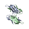
| |||||||||
|---|---|---|---|---|---|---|---|---|---|---|
| 1 |
| |||||||||
| NMR ensembles |
|
-Components
| #1: Protein | Mass: 11022.090 Da / Num. of mol.: 2 Source method: isolated from a genetically manipulated source Source: (gene. exp.) Nostoc ellipsosporum (bacteria) / Plasmid: pET / Species (production host): Escherichia coli / Production host: Has protein modification | Y | |
|---|
-Experimental details
-Experiment
| Experiment | Method: SOLUTION NMR |
|---|---|
| NMR experiment | Type: 2D IPAP [15N-1H]- HSQC HSQC |
| NMR details | Text: Residual dipolar couplings were measured in the presence of a colloidal phage solution of 11.5 mg/ml Pf1. |
- Sample preparation
| Details | Contents: 0.150 mM protein in 25 mM sodium phosphate buffer, pH 8.0 and 0.02 % NaN3 Solvent system: 90% H2O/10% D2O |
|---|---|
| Sample conditions | pH: 6 / Pressure: 1 atm / Temperature: 293 K |
-NMR measurement
| Radiation | Protocol: SINGLE WAVELENGTH / Monochromatic (M) / Laue (L): M |
|---|---|
| Radiation wavelength | Relative weight: 1 |
| NMR spectrometer | Type: Bruker DRX / Manufacturer: Bruker / Model: DRX / Field strength: 600 MHz |
- Processing
| NMR software |
| ||||||||||||||||
|---|---|---|---|---|---|---|---|---|---|---|---|---|---|---|---|---|---|
| Refinement | Method: Determination of the domain orientation for the solution structure of the dimer was carried out using a procedure analogous to the one described for determining the relative domain ...Method: Determination of the domain orientation for the solution structure of the dimer was carried out using a procedure analogous to the one described for determining the relative domain orientation in a two-domain protein fragment of a lectin. Software ordinal: 1 Details: The coordinates of the individual domains of the domain swapped dimer CV-N were taken directly from the X-Ray coordinates, 3EZM and 1L5B. The only protons added are the HNE1 of W(49/150) and ...Details: The coordinates of the individual domains of the domain swapped dimer CV-N were taken directly from the X-Ray coordinates, 3EZM and 1L5B. The only protons added are the HNE1 of W(49/150) and all the backbone amide protons (HN), since domain-domain orientation was based only on HN/HNE1 residual dipolar couplings. The starting coordinates were those of two pseudo-monomer units (AB' and A'B) extracted from the refined trigonal 1.5 X-ray structure, in which proline 51 at the junction between A and B was removed, allowing for free rotation around this junction. We then treated AB' and A'B as two independent sub-domains. Assuming that the orientation of the two sub-domains is fixed in solution (at least to a first approximation), the principal axis systems, or alignment frames, of sub-domains AB' and A'B should be equivalent to the alignment system of the entire molecule and, vice versa, to each other. Using the residual dipolar couplings we calculated the order tensor principal axis systems for each domain. Rotation of pseudo sub-domain A'B around the hinge at amino acid position 51 until a superposition of the individual coordinate frames was obtained yielded the final model of the solution dimer. | ||||||||||||||||
| NMR ensemble | Conformers submitted total number: 1 |
