 Movie
Movie Controller


+ Open data

- Basic information
| Entry | Database: PDB / ID: 1l5b | ||||||
|---|---|---|---|---|---|---|---|
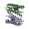
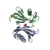
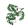
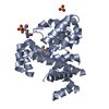
| Title | DOMAIN-SWAPPED CYANOVIRIN-N DIMER | ||||||
 Components Components | cyanovirin-N | ||||||
 Keywords Keywords | ANTIVIRAL PROTEIN / CYANOVIRIN-N / HIV-INACTIVATING / DOMAIN-SWAPPING / GP120 | ||||||
| Function / homology |  Function and homology information Function and homology informationoligosaccharide binding / regulation of defense response to virus / carbohydrate binding Similarity search - Function | ||||||
| Biological species |  Nostoc ellipsosporum (bacteria) Nostoc ellipsosporum (bacteria) | ||||||
| Method |  X-RAY DIFFRACTION / SYNCHROTRON / MOLECULAR REPLACEMENT / Resolution: 2 Å X-RAY DIFFRACTION / SYNCHROTRON / MOLECULAR REPLACEMENT / Resolution: 2 Å | ||||||
 Authors Authors | Barrientos, L.G. / Louis, J.M. / Botos, I. / Mori, T. / Han, Z. / O'Keefe, B.R. / Boyd, M.R. / Wlodawer, A. / Gronenborn, A.M. | ||||||
 Citation Citation | Journal: Structure / Year: 2002 Title: The domain-swapped dimer of cyanovirin-N is in a metastable folded state: reconciliation of X-ray and NMR structures. Authors: Barrientos, L.G. / Louis, J.M. / Botos, I. / Mori, T. / Han, Z. / O'Keefe, B.R. / Boyd, M.R. / Wlodawer, A. / Gronenborn, A.M. | ||||||
| History |
|
- Structure visualization
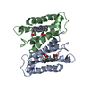
| Structure viewer | Molecule: MolmilJmol/JSmol |
|---|
- Downloads & links
-Download
| PDBx/mmCIF format | 1l5b.cif.gz | 58.1 KB | Display | PDBx/mmCIF format |
|---|---|---|---|---|
| PDB format | pdb1l5b.ent.gz | 41.6 KB | Display | PDB format |
| PDBx/mmJSON format | 1l5b.json.gz | Tree view | PDBx/mmJSON format | |
| Others |  Other downloads Other downloads |
-Validation report
| Arichive directory | https://data.pdbj.org/pub/pdb/validation_reports/l5/1l5bftp://data.pdbj.org/pub/pdb/validation_reports/l5/1l5b | HTTPS FTP |
|---|
-Related structure data
| Related structure data |  1l5eC  3ezmS S: Starting model for refinement C: citing same article ( |
|---|---|
| Similar structure data |


-Links
 PDBj
PDBj- Assembly
| Deposited unit | 
| ||||||||
|---|---|---|---|---|---|---|---|---|---|
| 1 |
| ||||||||
| Unit cell |
|
-Components
| #1: Protein | Mass: 11022.090 Da / Num. of mol.: 2 Source method: isolated from a genetically manipulated source Source: (gene. exp.) Nostoc ellipsosporum (bacteria) / Production host: #2: Chemical | ChemComp-NA / |   Mass: 22.990 Da / Num. of mol.: 1 / Source method: obtained synthetically / Formula: Na Mass: 22.990 Da / Num. of mol.: 1 / Source method: obtained synthetically / Formula: Na#3: Chemical | ChemComp-NHE / |   Mass: 207.290 Da / Num. of mol.: 1 / Source method: obtained synthetically / Formula: C8H17NO3S / Comment: pH buffer*YM Mass: 207.290 Da / Num. of mol.: 1 / Source method: obtained synthetically / Formula: C8H17NO3S / Comment: pH buffer*YM#4: Water | ChemComp-HOH / |  Mass: 18.015 Da / Num. of mol.: 195 / Source method: isolated from a natural source / Formula: H2O Mass: 18.015 Da / Num. of mol.: 195 / Source method: isolated from a natural source / Formula: H2OHas protein modification | Y | |
|---|
-Experimental details
-Experiment
| Experiment | Method: X-RAY DIFFRACTION / Number of used crystals: 1 |
|---|
- Sample preparation
| Crystal | Density Matthews: 3.23 Å3/Da / Density % sol: 61.93 % |
|---|---|
| Crystal grow | Temperature: 295 K / Method: vapor diffusion, hanging drop / pH: 10.3 Details: sodium citrate, CHES, pH 10.3, VAPOR DIFFUSION, HANGING DROP, temperature 295K |
-Data collection
| Diffraction | Mean temperature: 100 K |
|---|---|
| Diffraction source | Source: SYNCHROTRON / Site: NSLS  / Beamline: X9B / Wavelength: 0.97 Å / Beamline: X9B / Wavelength: 0.97 Å |
| Detector | Type: ADSC QUANTUM 4 / Detector: CCD / Date: Mar 4, 2001 |
| Radiation | Monochromator: SAGITALLY FOCUSED SI(111) / Protocol: SINGLE WAVELENGTH / Monochromatic (M) / Laue (L): M / Scattering type: x-ray |
| Radiation wavelength | Wavelength: 0.97 Å / Relative weight: 1 |
| Reflection | Resolution: 2→20 Å / Num. all: 16831 / Num. obs: 15588 / % possible obs: 76.7 % / Observed criterion σ(F): 0 / Observed criterion σ(I): -3 / Biso Wilson estimate: 22.4 Å2 / Rmerge(I) obs: 0.049 |
| Reflection shell | Resolution: 2→2.13 Å / % possible all: 87.8 |
| Reflection | *PLUS Highest resolution: 2.07 Å / Num. obs: 20437 / % possible obs: 99.4 % / Num. measured all: 392421 |
| Reflection shell | *PLUS Lowest resolution: 2.07 Å / % possible obs: 99.5 % |
- Processing
| Software |
| ||||||||||||||||||||||||||||||||||||
|---|---|---|---|---|---|---|---|---|---|---|---|---|---|---|---|---|---|---|---|---|---|---|---|---|---|---|---|---|---|---|---|---|---|---|---|---|---|
| Refinement | Method to determine structure: MOLECULAR REPLACEMENT Starting model: PDB ENTRY 3EZM Resolution: 2→19.9 Å / Rfactor Rfree error: 0.007 / Data cutoff high absF: 1076002.84 / Data cutoff low absF: 0 / Isotropic thermal model: RESTRAINED / Cross valid method: THROUGHOUT / σ(F): 0 / Stereochemistry target values: Engh & Huber
| ||||||||||||||||||||||||||||||||||||
| Solvent computation | Solvent model: FLAT MODEL / Bsol: 72.2724 Å2 / ksol: 0.342024 e/Å3 | ||||||||||||||||||||||||||||||||||||
| Displacement parameters | Biso mean: 43.4 Å2
| ||||||||||||||||||||||||||||||||||||
| Refine analyze |
| ||||||||||||||||||||||||||||||||||||
| Refinement step | Cycle: LAST / Resolution: 2→19.9 Å
| ||||||||||||||||||||||||||||||||||||
| Refine LS restraints |
| ||||||||||||||||||||||||||||||||||||
| LS refinement shell | Resolution: 2→2.13 Å / Rfactor Rfree error: 0.024 / Total num. of bins used: 6
| ||||||||||||||||||||||||||||||||||||
| Xplor file |
| ||||||||||||||||||||||||||||||||||||
| Refinement | *PLUS Rfactor obs: 0.237 / Rfactor Rfree: 0.245 / Rfactor Rwork: 0.237 / Highest resolution: 2.07 Å / Lowest resolution: 20 Å | ||||||||||||||||||||||||||||||||||||
| Solvent computation | *PLUS | ||||||||||||||||||||||||||||||||||||
| Displacement parameters | *PLUS | ||||||||||||||||||||||||||||||||||||
| Refine LS restraints | *PLUS
| ||||||||||||||||||||||||||||||||||||
| LS refinement shell | *PLUS Rfactor obs: 0.406 |
