 Movie
Movie Controller

[English] 日本語
 Yorodumi
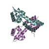
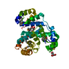
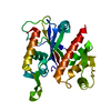
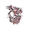
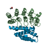
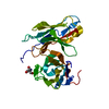
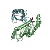
- PDB-2ccy: STRUCTURE OF FERRICYTOCHROME C(PRIME) FROM RHODOSPIRILLUM MOLISCH... -
+ Open data

- Basic information
| Entry | Database: PDB / ID: 2ccy | ||||||||||||
|---|---|---|---|---|---|---|---|---|---|---|---|---|---|
| Title | STRUCTURE OF FERRICYTOCHROME C(PRIME) FROM RHODOSPIRILLUM MOLISCHIANUM AT 1.67 ANGSTROMS RESOLUTION | ||||||||||||
 Components Components | CYTOCHROME C | ||||||||||||
 Keywords Keywords | ELECTRON TRANSPORT (HEME PROTEIN) | ||||||||||||
| Function / homology |  Function and homology information Function and homology informationelectron transport chain / electron transfer activity / periplasmic space / iron ion binding / heme binding Similarity search - Function | ||||||||||||
| Biological species |  Phaeospirillum molischianum (magnetotactic) Phaeospirillum molischianum (magnetotactic) | ||||||||||||
| Method |  X-RAY DIFFRACTION / Resolution: 1.67 Å X-RAY DIFFRACTION / Resolution: 1.67 Å | ||||||||||||
 Authors Authors | Finzel, B.C. / Weber, P.C. / Hardman, K.D. / Salemme, F.R. | ||||||||||||
 Citation Citation | Journal: J.Mol.Biol. / Year: 1985 Title: Structure of ferricytochrome c' from Rhodospirillum molischianum at 1.67 A resolution. Authors: Finzel, B.C. / Weber, P.C. / Hardman, K.D. / Salemme, F.R. #1: Journal: Nature / Year: 1985Title: Lattice Mobility and Anomalous Temperature Factor Behaviour in Cytochrome C(Prime) Authors: Finzel, B.C. / Salemme, F.R. #2: Journal: J.Mol.Biol. / Year: 1981Title: Crystallographic Structure of Rhodospirillum Molischianum Ferricytochrome C(Prime) at 2.5 Angstroms Resolution Authors: Weber, P.C. / Howard, A. / Xuong, N.H. / Salemme, F.R. #3: Journal: Biochemistry / Year: 1982Title: Correlations between Structural and Spectroscopic Properties of the High-Spin Heme Protein Cytochrome C(Prime) Authors: Weber, P.C. #4: Journal: J.Biol.Chem. / Year: 1981Title: On the Evolutionary Relationship of the 4-Alpha-Helical Heme Proteins. The Comparison of Cytochrome B562 and Cytochrome C(Prime) Authors: Weber, P.C. / Salemme, F.R. / Mathews, F.S. / Bethge, P.H. #5: Journal: Nature / Year: 1980Title: Structural and Functional Diversity in 4-Alpha-Helical Proteins Authors: Weber, P.C. / Salemme, F.R. #6: Journal: Nature / Year: 1980Title: Structure of Cytochrome C(Prime). A Dimeric, High-Spin Haem Protein Authors: Weber, P.C. / Bartsch, R.G. / Cusanovich, M.A. / Hamlin, R.C. / Howard, A. / Jordan, S.R. / Kamen, M.D. / Meyer, T.E. / Weatherford, D.W. / Xuong, N.H. / Salemme, F.R. #7: Journal: J.Mol.Biol. / Year: 1977Title: Preliminary Crystallographic Data for Cytochromes C(Prime) of Rhodopseudomonas Capsulata and Rhodospirillum Molischianum Authors: Weber, P. / Salemme, F.R. | ||||||||||||
| History |
|
- Structure visualization
| Structure viewer | Molecule: MolmilJmol/JSmol |
|---|
- Downloads & links
-Download
| PDBx/mmCIF format | 2ccy.cif.gz | 67.1 KB | Display | PDBx/mmCIF format |
|---|---|---|---|---|
| PDB format | pdb2ccy.ent.gz | 49.7 KB | Display | PDB format |
| PDBx/mmJSON format | 2ccy.json.gz | Tree view | PDBx/mmJSON format | |
| Others |  Other downloads Other downloads |
-Validation report
| Arichive directory | https://data.pdbj.org/pub/pdb/validation_reports/cc/2ccyftp://data.pdbj.org/pub/pdb/validation_reports/cc/2ccy | HTTPS FTP |
|---|
-Related structure data
| Similar structure data |
|---|


-Links
 PDBj
PDBj


- Assembly
| Deposited unit | 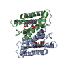
| ||||||||
|---|---|---|---|---|---|---|---|---|---|
| 1 |
| ||||||||
| Unit cell |
| ||||||||
| Atom site foot note | 1: RESIDUE GLN 42 IN BOTH CHAINS IS MODELLED IN TWO CONFORMATIONS. | ||||||||
| Noncrystallographic symmetry (NCS) | NCS oper: (Code: given Matrix: (0.4673, 0.2938, -0.8339), Vector: |
-Components
| #1: Protein | Mass: 13439.367 Da / Num. of mol.: 2 Source method: isolated from a genetically manipulated source Source: (gene. exp.) Phaeospirillum molischianum (magnetotactic)References: UniProt: P00152 #2: Chemical |   Mass: 618.503 Da / Num. of mol.: 2 / Source method: obtained synthetically / Formula: C34H34FeN4O4 Mass: 618.503 Da / Num. of mol.: 2 / Source method: obtained synthetically / Formula: C34H34FeN4O4#3: Water | ChemComp-HOH / |  Mass: 18.015 Da / Num. of mol.: 194 / Source method: isolated from a natural source / Formula: H2O Mass: 18.015 Da / Num. of mol.: 194 / Source method: isolated from a natural source / Formula: H2OHas protein modification | Y | |
|---|
-Experimental details
-Experiment
| Experiment | Method: X-RAY DIFFRACTION |
|---|
- Sample preparation
| Crystal | Density Matthews: 2.87 Å3/Da / Density % sol: 57.14 % | |||||||||||||||||||||||||
|---|---|---|---|---|---|---|---|---|---|---|---|---|---|---|---|---|---|---|---|---|---|---|---|---|---|---|
| Crystal grow | *PLUS Temperature: 4 ℃ / Method: vapor diffusionDetails: referred to 'Weber, P.', (1977) J.Mol.Biol., 117, 815-820 | |||||||||||||||||||||||||
| Components of the solutions | *PLUS
|
-Data collection
| Reflection | *PLUS Highest resolution: 1.5 Å / Lowest resolution: 1.66 Å / Num. obs: 46290 / % possible obs: 72 % / Observed criterion σ(I): 1 / Num. measured all: 213471 |
|---|
- Processing
| Software | Name: PROLSQ / Classification: refinement | ||||||||||||||||||||||||||||||||||||||||||||||||||||||||||||
|---|---|---|---|---|---|---|---|---|---|---|---|---|---|---|---|---|---|---|---|---|---|---|---|---|---|---|---|---|---|---|---|---|---|---|---|---|---|---|---|---|---|---|---|---|---|---|---|---|---|---|---|---|---|---|---|---|---|---|---|---|---|
| Refinement | Highest resolution: 1.67 Å / Num. reflection obs: 30533 / σ(I): 1 | ||||||||||||||||||||||||||||||||||||||||||||||||||||||||||||
| Refinement step | Cycle: LAST / Highest resolution: 1.67 Å
| ||||||||||||||||||||||||||||||||||||||||||||||||||||||||||||
| Refine LS restraints |
| ||||||||||||||||||||||||||||||||||||||||||||||||||||||||||||
| Refinement | *PLUS σ(I): 1 / Highest resolution: 1.67 Å / Lowest resolution: 9999 Å / Num. reflection obs: 30533 / Rfactor obs: 0.188 | ||||||||||||||||||||||||||||||||||||||||||||||||||||||||||||
| Solvent computation | *PLUS | ||||||||||||||||||||||||||||||||||||||||||||||||||||||||||||
| Displacement parameters | *PLUS | ||||||||||||||||||||||||||||||||||||||||||||||||||||||||||||
| Refine LS restraints | *PLUS
| ||||||||||||||||||||||||||||||||||||||||||||||||||||||||||||
| LS refinement shell | *PLUS Lowest resolution: 9999 Å |
