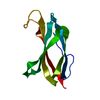
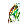
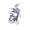
Journal: J Mol Biol / Year: 2007 Title: Crystal structure of human filamin C domain 23 and small angle scattering model for filamin C 23-24 dimer. Authors: Ljiljana Sjekloća / Regina Pudas / Björn Sjöblom / Peter Konarev / Oliviero Carugo / Vladimir Rybin / Tiila-Riikka Kiema / Dmitri Svergun / Jari Ylänne / Kristina Djinović Carugo / Abstract: Filamin C is a dimeric, actin-binding protein involved in organization of cortical cytoskeleton and of the sarcomere. We performed crystallographic, small-angle X-ray scattering and analytical ...Filamin C is a dimeric, actin-binding protein involved in organization of cortical cytoskeleton and of the sarcomere. We performed crystallographic, small-angle X-ray scattering and analytical ultracentrifugation experiments on the constructs containing carboxy-terminal domains of the protein (domains 23-24 and 19-21). The crystal structure of domain 23 of filamin C showed that the protein adopts the expected immunoglobulin (Ig)-like fold. Small-angle X-ray scattering experiments performed on filamin C tandem Ig-like domains 23 and 24 reveal a dimer that is formed by domain 24 and that domain 23 has little interactions with itself or with domain 24, while the analytical ultracentrifugation experiments showed that the filamin C domains 19-21 form elongated monomers in diluted solutions.
History
Deposition
Oct 31, 2006
Deposition site: RCSB / Processing site: PDBJ
Revision 1.0
Sep 11, 2007
Provider: repository / Type: Initial release
Revision 1.1
Jul 13, 2011
Group: Non-polymer description / Version format compliance
Resolution: 2.05→2.16 Å / Redundancy: 6.5 % / Rmerge(I) obs: 0.298 / Mean I/σ(I) obs: 2.1 / Num. unique all: 950 / Rsym value: 0.298 / % possible all: 97.7
-
Processing
Software
Name
Version
Classification
NB
REFMAC
5.2.0005
refinement
PDB_EXTRACT
2
dataextraction
MAR345
datacollection
DENZO
datareduction
SCALA
datascaling
SHARP
phasing
Refinement
Method to determine structure: SAD / Resolution: 2.05→45.17 Å / Cor.coef. Fo:Fc: 0.96 / Cor.coef. Fo:Fc free: 0.935 / SU B: 3.855 / SU ML: 0.108 / Cross valid method: THROUGHOUT / σ(F): 0 / ESU R: 0.179 / ESU R Free: 0.168 / Stereochemistry target values: MAXIMUM LIKELIHOOD / Details: HYDROGENS HAVE BEEN ADDED IN THE RIDING POSITIONS
Rfactor
Num. reflection
% reflection
Selection details
Rfree
0.237
329
4.7 %
RANDOM
Rwork
0.188
-
-
-
all
0.19
7116
-
-
obs
0.19
7016
97.51 %
-
Solvent computation
Ion probe radii: 0.8 Å / Shrinkage radii: 0.8 Å / VDW probe radii: 1.2 Å / Solvent model: BABINET MODEL WITH MASK
Displacement parameters
Biso mean: 36.963 Å2
Baniso -1
Baniso -2
Baniso -3
1-
0.55 Å2
0.28 Å2
0 Å2
2-
-
0.55 Å2
0 Å2
3-
-
-
-0.83 Å2
Refinement step
Cycle: LAST / Resolution: 2.05→45.17 Å
Protein
Nucleic acid
Ligand
Solvent
Total
Num. atoms
689
0
23
56
768
Refine LS restraints
Refine-ID
Type
Dev ideal
Dev ideal target
Number
X-RAY DIFFRACTION
r_bond_refined_d
0.012
0.022
712
X-RAY DIFFRACTION
r_bond_other_d
0.004
0.02
651
X-RAY DIFFRACTION
r_angle_refined_deg
1.288
2.018
966
X-RAY DIFFRACTION
r_angle_other_deg
0.728
3
1519
X-RAY DIFFRACTION
r_dihedral_angle_1_deg
5.543
5
96
X-RAY DIFFRACTION
r_dihedral_angle_2_deg
37.058
24.737
19
X-RAY DIFFRACTION
r_dihedral_angle_3_deg
13.785
15
95
X-RAY DIFFRACTION
r_dihedral_angle_4_deg
7.013
15
1
X-RAY DIFFRACTION
r_chiral_restr
0.073
0.2
109
X-RAY DIFFRACTION
r_gen_planes_refined
0.005
0.02
798
X-RAY DIFFRACTION
r_gen_planes_other
0.001
0.02
131
X-RAY DIFFRACTION
r_nbd_refined
0.181
0.2
85
X-RAY DIFFRACTION
r_nbd_other
0.229
0.2
539
X-RAY DIFFRACTION
r_nbtor_refined
0.173
0.2
323
X-RAY DIFFRACTION
r_nbtor_other
0.089
0.2
403
X-RAY DIFFRACTION
r_xyhbond_nbd_refined
0.113
0.2
34
X-RAY DIFFRACTION
r_metal_ion_refined
0.026
0.2
1
X-RAY DIFFRACTION
r_symmetry_vdw_refined
0.11
0.2
4
X-RAY DIFFRACTION
r_symmetry_vdw_other
0.259
0.2
19
X-RAY DIFFRACTION
r_symmetry_hbond_refined
0.098
0.2
8
X-RAY DIFFRACTION
r_mcbond_it
1.34
1.5
605
X-RAY DIFFRACTION
r_mcbond_other
0.191
1.5
203
X-RAY DIFFRACTION
r_mcangle_it
1.554
2
766
X-RAY DIFFRACTION
r_scbond_it
2.379
3
267
X-RAY DIFFRACTION
r_scangle_it
3.207
4.5
200
LS refinement shell
Resolution: 2.05→2.104 Å / Total num. of bins used: 20
Rfactor
Num. reflection
% reflection
Rfree
0.219
19
-
Rwork
0.213
485
-
obs
-
504
94.38 %
+
About Yorodumi
-
News
-
Feb 9, 2022. New format data for meta-information of EMDB entries
New format data for meta-information of EMDB entries
Version 3 of the EMDB header file is now the official format.
The previous official version 1.9 will be removed from the archive.
In the structure databanks used in Yorodumi, some data are registered as the other names, "COVID-19 virus" and "2019-nCoV". Here are the details of the virus and the list of structure data.
Jan 31, 2019. EMDB accession codes are about to change! (news from PDBe EMDB page)
EMDB accession codes are about to change! (news from PDBe EMDB page)
The allocation of 4 digits for EMDB accession codes will soon come to an end. Whilst these codes will remain in use, new EMDB accession codes will include an additional digit and will expand incrementally as the available range of codes is exhausted. The current 4-digit format prefixed with “EMD-” (i.e. EMD-XXXX) will advance to a 5-digit format (i.e. EMD-XXXXX), and so on. It is currently estimated that the 4-digit codes will be depleted around Spring 2019, at which point the 5-digit format will come into force.
The EM Navigator/Yorodumi systems omit the EMD- prefix.
Related info.:Q: What is EMD? / ID/Accession-code notation in Yorodumi/EM Navigator
Yorodumi is a browser for structure data from EMDB, PDB, SASBDB, etc.
This page is also the successor to EM Navigator detail page, and also detail information page/front-end page for Omokage search.
The word "yorodu" (or yorozu) is an old Japanese word meaning "ten thousand". "mi" (miru) is to see.
Related info.:EMDB / PDB / SASBDB / Comparison of 3 databanks / Yorodumi Search / Aug 31, 2016. New EM Navigator & Yorodumi / Yorodumi Papers / Jmol/JSmol / Function and homology information / Changes in new EM Navigator and Yorodumi
 Movie
Movie Controller


 Open data
 Basic information
 Components
 Keywords
 Function and homology information
 Homo sapiens (human)
 X-RAY DIFFRACTION /
  Authors
 Citation
 Structure visualization
 Downloads & links
 Other downloads


 PDBj
PDBj


 Assembly
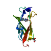


 Mass: 58.693 Da / Num. of mol.: 1 / Source method: obtained synthetically / Formula: Ni
 Mass: 69.085 Da / Num. of mol.: 2 / Source method: obtained synthetically / Formula: C3H5N2
 Mass: 92.094 Da / Num. of mol.: 2 / Source method: obtained synthetically / Formula: C3H8O3
 Mass: 18.015 Da / Num. of mol.: 56 / Source method: isolated from a natural source / Formula: H2O
 Sample preparation
 Processing