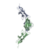
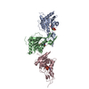
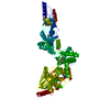
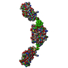
Filamin C 23-24 (protein), FilaminC 23-24, Escherichia coli
Biological species
Escherichia coli (E. coli)
Citation
Journal: J Mol Biol / Year: 2007 Title: Crystal structure of human filamin C domain 23 and small angle scattering model for filamin C 23-24 dimer. Authors: Ljiljana Sjekloća / Regina Pudas / Björn Sjöblom / Peter Konarev / Oliviero Carugo / Vladimir Rybin / Tiila-Riikka Kiema / Dmitri Svergun / Jari Ylänne / Kristina Djinović Carugo / Abstract: Filamin C is a dimeric, actin-binding protein involved in organization of cortical cytoskeleton and of the sarcomere. We performed crystallographic, small-angle X-ray scattering and analytical ...Filamin C is a dimeric, actin-binding protein involved in organization of cortical cytoskeleton and of the sarcomere. We performed crystallographic, small-angle X-ray scattering and analytical ultracentrifugation experiments on the constructs containing carboxy-terminal domains of the protein (domains 23-24 and 19-21). The crystal structure of domain 23 of filamin C showed that the protein adopts the expected immunoglobulin (Ig)-like fold. Small-angle X-ray scattering experiments performed on filamin C tandem Ig-like domains 23 and 24 reveal a dimer that is formed by domain 24 and that domain 23 has little interactions with itself or with domain 24, while the analytical ultracentrifugation experiments showed that the filamin C domains 19-21 form elongated monomers in diluted solutions.
Contact author
Petr Konarev (EMBL-Hamburg, European Molecular Biology Laboratory (EMBL) - Hamburg outstation, Notkestraße 85, Geb. 25A, 22607 Hamburg, Deutschland, Germany)
Instrument name: DORIS III X33 / City: Hamburg / 国: Germany / Type of source: X-ray synchrotron
Detector
Name: MAR 345 Image Plate
Scan
Title: Filamin C 23-24 / Measurement date: Jun 17, 2005 / Storage temperature: 10 °C / Cell temperature: 10 °C / Exposure time: 120 sec. / Number of frames: 2 / Unit: 1/nm /
Min
Max
Q
0.1893
4.5815
Distance distribution function P(R)
Sofotware P(R): GNOM 4.5a / Number of points: 480 /
Min
Max
Q
0.1916
4.581
P(R) point
10
489
R
0
13
Result
Type of curve: merged /
Experimental
Porod
MW
42 kDa
-
Volume
-
75 nm3
P(R)
Guinier
Forward scattering, I0
121
122
Radius of gyration, Rg
3.53 nm
3.5 nm
Min
Max
D
-
13
Guinier point
1
25
+
About Yorodumi
-
News
-
Feb 9, 2022. New format data for meta-information of EMDB entries
New format data for meta-information of EMDB entries
Version 3 of the EMDB header file is now the official format.
The previous official version 1.9 will be removed from the archive.
In the structure databanks used in Yorodumi, some data are registered as the other names, "COVID-19 virus" and "2019-nCoV". Here are the details of the virus and the list of structure data.
Jan 31, 2019. EMDB accession codes are about to change! (news from PDBe EMDB page)
EMDB accession codes are about to change! (news from PDBe EMDB page)
The allocation of 4 digits for EMDB accession codes will soon come to an end. Whilst these codes will remain in use, new EMDB accession codes will include an additional digit and will expand incrementally as the available range of codes is exhausted. The current 4-digit format prefixed with “EMD-” (i.e. EMD-XXXX) will advance to a 5-digit format (i.e. EMD-XXXXX), and so on. It is currently estimated that the 4-digit codes will be depleted around Spring 2019, at which point the 5-digit format will come into force.
The EM Navigator/Yorodumi systems omit the EMD- prefix.
Related info.:Q: What is EMD? / ID/Accession-code notation in Yorodumi/EM Navigator
Yorodumi is a browser for structure data from EMDB, PDB, SASBDB, etc.
This page is also the successor to EM Navigator detail page, and also detail information page/front-end page for Omokage search.
The word "yorodu" (or yorozu) is an old Japanese word meaning "ten thousand". "mi" (miru) is to see.
Related info.:EMDB / PDB / SASBDB / Comparison of 3 databanks / Yorodumi Search / Aug 31, 2016. New EM Navigator & Yorodumi / Yorodumi Papers / Jmol/JSmol / Function and homology information / Changes in new EM Navigator and Yorodumi
 Movie
Movie Controller

 Open data
 Basic information
 Sample
 Citation
 Contact author
 Structure visualization
 Molmil
 Downloads & links
 SASDAC4
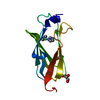


 Search similar-shape structures of this assembly by Omokage search (details)
 DORIS III X33
  / City: Hamburg / 国: Germany


