[English] 日本語
Yorodumi
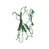
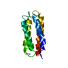
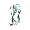
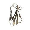
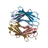
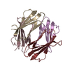
- PDB-6msv: Structure of the 6th type III domain from human fibronectin -

+
Open data


ID or keywords:

Loading...

-
Basic information

Entry
Database: PDB / ID: 6msv
TitleStructure of the 6th type III domain from human fibronectin
ComponentsFibronectin
KeywordsCELL ADHESION
Function / homology
Function and homology information


negative regulation of monocyte activation / calcium-independent cell-matrix adhesion / negative regulation of transforming growth factor beta production / Fibronectin matrix formation / Extracellular matrix organization / positive regulation of substrate-dependent cell migration, cell attachment to substrate / neural crest cell migration involved in autonomic nervous system development / peptidase activator activity / fibrinogen complex / peptide cross-linking ...negative regulation of monocyte activation / calcium-independent cell-matrix adhesion / negative regulation of transforming growth factor beta production / Fibronectin matrix formation / Extracellular matrix organization / positive regulation of substrate-dependent cell migration, cell attachment to substrate / neural crest cell migration involved in autonomic nervous system development / peptidase activator activity / fibrinogen complex / peptide cross-linking / integrin activation / ALK mutants bind TKIs / cell-substrate junction assembly / biological process involved in interaction with symbiont / proteoglycan binding / Molecules associated with elastic fibres / MET activates PTK2 signaling / extracellular matrix structural constituent / Syndecan interactions / p130Cas linkage to MAPK signaling for integrins / endodermal cell differentiation / GRB2:SOS provides linkage to MAPK signaling for Integrins / endoplasmic reticulum-Golgi intermediate compartment / Non-integrin membrane-ECM interactions / basement membrane / ECM proteoglycans / Integrin cell surface interactions / positive regulation of axon extension / collagen binding / Degradation of the extracellular matrix / Integrin signaling / substrate adhesion-dependent cell spreading / regulation of ERK1 and ERK2 cascade / cell-matrix adhesion / extracellular matrix / platelet alpha granule lumen / acute-phase response / integrin-mediated signaling pathway / Cell surface interactions at the vascular wall / Post-translational protein phosphorylation / regulation of protein phosphorylation / Signaling by high-kinase activity BRAF mutants / wound healing / MAP2K and MAPK activation / response to wounding / Signaling by RAF1 mutants / Signaling by moderate kinase activity BRAF mutants / Paradoxical activation of RAF signaling by kinase inactive BRAF / GPER1 signaling / Signaling downstream of RAS mutants / Regulation of Insulin-like Growth Factor (IGF) transport and uptake by Insulin-like Growth Factor Binding Proteins (IGFBPs) / Signaling by BRAF and RAF1 fusions / positive regulation of fibroblast proliferation / Signaling by ALK fusions and activated point mutants / integrin binding / Platelet degranulation / nervous system development / heparin binding / heart development / regulation of cell shape / angiogenesis / Interleukin-4 and Interleukin-13 signaling / collagen-containing extracellular matrix / protease binding / positive regulation of phosphatidylinositol 3-kinase/protein kinase B signal transduction / blood microparticle / cell adhesion / apical plasma membrane / endoplasmic reticulum lumen / signaling receptor binding / positive regulation of cell population proliferation / positive regulation of gene expression / extracellular space / extracellular exosome / extracellular region / identical protein binding / plasma membrane
Similarity search - Function
Fibronectin type I domain / Fibronectin, type I / Fibronectin type-I domain signature. / Fibronectin type-I domain profile. / Fibronectin type 1 domain / Fibronectin type II domain / Fibronectin type II domain superfamily / Fibronectin type II domain / Fibronectin type-II collagen-binding domain signature. / Fibronectin type-II collagen-binding domain profile. ...Fibronectin type I domain / Fibronectin, type I / Fibronectin type-I domain signature. / Fibronectin type-I domain profile. / Fibronectin type 1 domain / Fibronectin type II domain / Fibronectin type II domain superfamily / Fibronectin type II domain / Fibronectin type-II collagen-binding domain signature. / Fibronectin type-II collagen-binding domain profile. / Fibronectin type 2 domain / Kringle-like fold / Fibronectin type III domain / EGF-like domain signature 1. / Fibronectin type 3 domain / Fibronectin type-III domain profile. / Fibronectin type III / Fibronectin type III superfamily / Immunoglobulin-like fold
Similarity search - Domain/homology
Biological speciesHomo sapiens (human)
MethodX-RAY DIFFRACTION / SYNCHROTRON / MOLECULAR REPLACEMENT / Resolution: 2.4 Å
AuthorsLoa, S. / Mou, T.C. / Sprang, S.R. / Briknarova, K.
Funding support United States, 2items
OrganizationGrant numberCountry
National Institutes of Health/National Institute of General Medical Sciences (NIH/NIGMS)R01GM114657 United States
National Institutes of Health/National Institute of General Medical Sciences (NIH/NIGMS)P20GM103546 United States
CitationJournal: To Be Published
Title: Structure of the 6th type III domain from human fibronectin
Authors: Loa, S. / Mou, T.C. / Sprang, S.R. / Briknarova, K.
History
DepositionOct 18, 2018Deposition site: RCSB / Processing site: RCSB
Revision 1.0Oct 23, 2019Provider: repository / Type: Initial release
Revision 1.1Jan 1, 2020Group: Author supporting evidence / Category: pdbx_audit_support / Item: _pdbx_audit_support.funding_organization
Revision 1.2Oct 11, 2023Group: Data collection / Database references / Refinement description
Category: chem_comp_atom / chem_comp_bond ...chem_comp_atom / chem_comp_bond / database_2 / pdbx_initial_refinement_model
Item: _database_2.pdbx_DOI / _database_2.pdbx_database_accession

-
Structure visualization

Structure viewerMolecule:
MolmilJmol/JSmol

Downloads & links

-
Assembly

Deposited unit
A: Fibronectin
B: Fibronectin
C: Fibronectin
D: Fibronectin
E: Fibronectin
F: Fibronectin
G: Fibronectin
H: Fibronectin
I: Fibronectin
J: Fibronectin
K: Fibronectin
L: Fibronectin
hetero molecules


Theoretical massNumber of molelcules
Total (without water)116,58214
Polymers116,39812
Non-polymers1842
Water2,666148
1
A: Fibronectin


Theoretical massNumber of molelcules
Total (without water)9,7001
Polymers9,7001
Non-polymers00
Water181
TypeNameSymmetry operationNumber
identity operation1_555x,y,z1
2
B: Fibronectin
hetero molecules


Theoretical massNumber of molelcules
Total (without water)9,7922
Polymers9,7001
Non-polymers921
Water181
TypeNameSymmetry operationNumber
identity operation1_555x,y,z1
3
C: Fibronectin


Theoretical massNumber of molelcules
Total (without water)9,7001
Polymers9,7001
Non-polymers00
Water181
TypeNameSymmetry operationNumber
identity operation1_555x,y,z1
4
D: Fibronectin


Theoretical massNumber of molelcules
Total (without water)9,7001
Polymers9,7001
Non-polymers00
Water181
TypeNameSymmetry operationNumber
identity operation1_555x,y,z1
5
E: Fibronectin


Theoretical massNumber of molelcules
Total (without water)9,7001
Polymers9,7001
Non-polymers00
Water181
TypeNameSymmetry operationNumber
identity operation1_555x,y,z1
6
F: Fibronectin
hetero molecules


Theoretical massNumber of molelcules
Total (without water)9,7922
Polymers9,7001
Non-polymers921
Water181
TypeNameSymmetry operationNumber
identity operation1_555x,y,z1
7
G: Fibronectin


Theoretical massNumber of molelcules
Total (without water)9,7001
Polymers9,7001
Non-polymers00
Water181
TypeNameSymmetry operationNumber
identity operation1_555x,y,z1
8
H: Fibronectin


Theoretical massNumber of molelcules
Total (without water)9,7001
Polymers9,7001
Non-polymers00
Water181
TypeNameSymmetry operationNumber
identity operation1_555x,y,z1
9
I: Fibronectin


Theoretical massNumber of molelcules
Total (without water)9,7001
Polymers9,7001
Non-polymers00
Water181
TypeNameSymmetry operationNumber
identity operation1_555x,y,z1
10
J: Fibronectin


Theoretical massNumber of molelcules
Total (without water)9,7001
Polymers9,7001
Non-polymers00
Water181
TypeNameSymmetry operationNumber
identity operation1_555x,y,z1
11
K: Fibronectin


Theoretical massNumber of molelcules
Total (without water)9,7001
Polymers9,7001
Non-polymers00
Water181
TypeNameSymmetry operationNumber
identity operation1_555x,y,z1
12
L: Fibronectin


Theoretical massNumber of molelcules
Total (without water)9,7001
Polymers9,7001
Non-polymers00
Water181
TypeNameSymmetry operationNumber
identity operation1_555x,y,z1
13
A: Fibronectin
G: Fibronectin
H: Fibronectin
J: Fibronectin


Theoretical massNumber of molelcules
Total (without water)38,7994
Polymers38,7994
Non-polymers00
Water724
TypeNameSymmetry operationNumber
identity operation1_555x,y,z1
Buried area5410 Å2
ΔGint-25 kcal/mol
Surface area14810 Å2
MethodPISA
14
B: Fibronectin
D: Fibronectin
F: Fibronectin
L: Fibronectin
hetero molecules


Theoretical massNumber of molelcules
Total (without water)38,9846
Polymers38,7994
Non-polymers1842
Water724
TypeNameSymmetry operationNumber
identity operation1_555x,y,z1
Buried area5230 Å2
ΔGint-22 kcal/mol
Surface area15170 Å2
MethodPISA
15
C: Fibronectin
E: Fibronectin
I: Fibronectin
K: Fibronectin


Theoretical massNumber of molelcules
Total (without water)38,7994
Polymers38,7994
Non-polymers00
Water724
TypeNameSymmetry operationNumber
identity operation1_555x,y,z1
Buried area5270 Å2
ΔGint-24 kcal/mol
Surface area14450 Å2
MethodPISA
Unit cell
Length a, b, c (Å)48.520, 79.460, 80.886
Angle α, β, γ (deg.)112.33, 95.88, 94.48
Int Tables number1
Space group name H-MP1

-
Components

#1: Protein
Fibronectin / FN / Cold-insoluble globulin / CIG


Mass: 9699.831 Da / Num. of mol.: 12
Source method: isolated from a genetically manipulated source
Source: (gene. exp.) Homo sapiens (human) / Gene: FN1, FN / Production host: Escherichia coli (E. coli) / References: UniProt: P02751
#2: Chemical ChemComp-GOL / GLYCEROL / GLYCERIN / PROPANE-1,2,3-TRIOL


Mass: 92.094 Da / Num. of mol.: 2 / Source method: obtained synthetically / Formula: C3H8O3
#3: Water ChemComp-HOH / water


Mass: 18.015 Da / Num. of mol.: 148 / Source method: isolated from a natural source / Formula: H2O

-
Experimental details

-
Experiment

ExperimentMethod: X-RAY DIFFRACTION / Number of used crystals: 1

-
Sample preparation

CrystalDensity Matthews: 2.48 Å3/Da / Density % sol: 50.44 %
Crystal growTemperature: 293 K / Method: vapor diffusion, hanging drop / Details: 2.0 M Ammonium Sulfate

-
Data collection

DiffractionMean temperature: 100 K / Serial crystal experiment: N
Diffraction sourceSource: SYNCHROTRON / Site: SSRL / Beamline: BL14-1 / Wavelength: 1.033 Å
DetectorType: MARMOSAIC 325 mm CCD / Detector: CCD / Date: May 7, 2017
RadiationProtocol: SINGLE WAVELENGTH / Monochromatic (M) / Laue (L): M / Scattering type: x-ray
Radiation wavelengthWavelength: 1.033 Å / Relative weight: 1
ReflectionResolution: 2.3→26.64 Å / Num. obs: 46905 / % possible obs: 90.2 % / Redundancy: 3.6 % / Biso Wilson estimate: 41.3 Å2 / Rsym value: 0.08 / Net I/σ(I): 13.1
Reflection shellResolution: 2.3→2.38 Å / Redundancy: 3.2 % / Mean I/σ(I) obs: 1.6 / Num. unique obs: 4387 / Rsym value: 0.66 / % possible all: 78.1

-
Processing

Software
NameVersionClassification
PHENIX(1.14_3260: ???)refinement
HKL-2000data reduction
HKL-2000data scaling
PHASERphasing
RefinementMethod to determine structure: MOLECULAR REPLACEMENT
Starting model: 5DFT
Resolution: 2.4→26.64 Å / SU ML: 0.44 / Cross valid method: FREE R-VALUE / σ(F): 0.1 / Phase error: 35.62
RfactorNum. reflection% reflection
Rfree0.295 1680 4.25 %
Rwork0.2353 --
obs0.2379 39570 91.83 %
Solvent computationShrinkage radii: 0.9 Å / VDW probe radii: 1.11 Å
Refinement stepCycle: LAST / Resolution: 2.4→26.64 Å
ProteinNucleic acidLigandSolventTotal
Num. atoms7667 0 12 148 7827
Refine LS restraints
Refine-IDTypeDev idealNumber
X-RAY DIFFRACTIONf_bond_d0.0047846
X-RAY DIFFRACTIONf_angle_d0.70210779
X-RAY DIFFRACTIONf_dihedral_angle_d9.4994749
X-RAY DIFFRACTIONf_chiral_restr0.0531303
X-RAY DIFFRACTIONf_plane_restr0.0061401
LS refinement shell
Resolution (Å)Rfactor RfreeNum. reflection RfreeRfactor RworkNum. reflection RworkRefine-ID% reflection obs (%)
2.4-2.47060.40061230.36352902X-RAY DIFFRACTION84
2.4706-2.55030.39121280.34082955X-RAY DIFFRACTION85
2.5503-2.64130.38371370.31782928X-RAY DIFFRACTION86
2.6413-2.7470.4041320.32582959X-RAY DIFFRACTION86
2.747-2.87190.34231390.29993122X-RAY DIFFRACTION90
2.8719-3.02310.4011440.27143188X-RAY DIFFRACTION93
3.0231-3.21220.30071420.24613245X-RAY DIFFRACTION94
3.2122-3.45970.31581460.24063298X-RAY DIFFRACTION96
3.4597-3.8070.27811450.21763285X-RAY DIFFRACTION97
3.807-4.35580.2611490.21873344X-RAY DIFFRACTION97
4.3558-5.480.26141460.18193323X-RAY DIFFRACTION97
5.48-26.64110.24361490.20533341X-RAY DIFFRACTION97

+
About Yorodumi

-
News

-
Feb 9, 2022. New format data for meta-information of EMDB entries

New format data for meta-information of EMDB entries

  • Version 3 of the EMDB header file is now the official format.
  • The previous official version 1.9 will be removed from the archive.

Related info.:EMDB header

External links:wwPDB to switch to version 3 of the EMDB data model

-
Aug 12, 2020. Covid-19 info

Covid-19 info

URL: https://pdbjlvh1.pdbj.org/emnavi/covid19.php

New page: Covid-19 featured information page in EM Navigator.

Related info.:Covid-19 info / Mar 5, 2020. Novel coronavirus structure data

+
Mar 5, 2020. Novel coronavirus structure data

Novel coronavirus structure data

Related info.:Yorodumi Speices / Aug 12, 2020. Covid-19 info

External links:COVID-19 featured content - PDBj / Molecule of the Month (242):Coronavirus Proteases

+
Jan 31, 2019. EMDB accession codes are about to change! (news from PDBe EMDB page)

EMDB accession codes are about to change! (news from PDBe EMDB page)

  • The allocation of 4 digits for EMDB accession codes will soon come to an end. Whilst these codes will remain in use, new EMDB accession codes will include an additional digit and will expand incrementally as the available range of codes is exhausted. The current 4-digit format prefixed with “EMD-” (i.e. EMD-XXXX) will advance to a 5-digit format (i.e. EMD-XXXXX), and so on. It is currently estimated that the 4-digit codes will be depleted around Spring 2019, at which point the 5-digit format will come into force.
  • The EM Navigator/Yorodumi systems omit the EMD- prefix.

Related info.:Q: What is EMD? / ID/Accession-code notation in Yorodumi/EM Navigator

External links:EMDB Accession Codes are Changing Soon! / Contact to PDBj

+
Jul 12, 2017. Major update of PDB

Major update of PDB

  • wwPDB released updated PDB data conforming to the new PDBx/mmCIF dictionary.
  • This is a major update changing the version number from 4 to 5, and with Remediation, in which all the entries are updated.
  • In this update, many items about electron microscopy experimental information are reorganized (e.g. em_software).
  • Now, EM Navigator and Yorodumi are based on the updated data.

External links:wwPDB Remediation / Enriched Model Files Conforming to OneDep Data Standards Now Available in the PDB FTP Archive

-
Yorodumi

Thousand views of thousand structures

  • Yorodumi is a browser for structure data from EMDB, PDB, SASBDB, etc.
  • This page is also the successor to EM Navigator detail page, and also detail information page/front-end page for Omokage search.
  • The word "yorodu" (or yorozu) is an old Japanese word meaning "ten thousand". "mi" (miru) is to see.

Related info.:EMDB / PDB / SASBDB / Comparison of 3 databanks / Yorodumi Search / Aug 31, 2016. New EM Navigator & Yorodumi / Yorodumi Papers / Jmol/JSmol / Function and homology information / Changes in new EM Navigator and Yorodumi

Read more