[English] 日本語
Yorodumi
- PDB-4wtx: Crystal structure of the fourth FnIII domain of integrin beta4 -

+
Open data


ID or keywords:

Loading...

-
Basic information

Entry
Database: PDB / ID: 4wtx
TitleCrystal structure of the fourth FnIII domain of integrin beta4
ComponentsIntegrin beta-4
KeywordsCELL ADHESION / FIBRONECTIN TYPE III / INTEGRIN / RECEPTOR
Function / homology
Function and homology information


trophoblast cell migration / Type I hemidesmosome assembly / hemidesmosome assembly / nail development / hemidesmosome / peripheral nervous system myelin formation / Laminin interactions / skin morphogenesis / filopodium assembly / mesodermal cell differentiation ...trophoblast cell migration / Type I hemidesmosome assembly / hemidesmosome assembly / nail development / hemidesmosome / peripheral nervous system myelin formation / Laminin interactions / skin morphogenesis / filopodium assembly / mesodermal cell differentiation / Differentiation of keratinocytes in interfollicular epidermis in mammalian skin / integrin complex / Assembly of collagen fibrils and other multimeric structures / Syndecan interactions / cell leading edge / basement membrane / cell-matrix adhesion / basal plasma membrane / integrin-mediated signaling pathway / G protein-coupled receptor binding / cell motility / cell-cell adhesion / response to wounding / autophagy / integrin binding / cell junction / nuclear membrane / receptor complex / cell adhesion / focal adhesion / nucleolus / cell surface / extracellular exosome / metal ion binding / plasma membrane
Similarity search - Function
Integrin beta-4 subunit / Na-Ca exchanger/integrin-beta4 / Calx-beta domain / Domains in Na-Ca exchangers and integrin-beta4 / CalX-like domain superfamily / Integrin beta, epidermal growth factor-like domain 1 / Integrin beta epidermal growth factor like domain 1 / Integrin beta tail domain / Integrin beta subunit, tail / Integrin beta tail domain superfamily ...Integrin beta-4 subunit / Na-Ca exchanger/integrin-beta4 / Calx-beta domain / Domains in Na-Ca exchangers and integrin-beta4 / CalX-like domain superfamily / Integrin beta, epidermal growth factor-like domain 1 / Integrin beta epidermal growth factor like domain 1 / Integrin beta tail domain / Integrin beta subunit, tail / Integrin beta tail domain superfamily / Integrin_B_tail / Integrin beta subunit, VWA domain / Integrin beta subunit / Integrin beta N-terminal / Integrin beta chain VWA domain / Integrin plexin domain / Integrins beta chain cysteine-rich domain signature. / Integrin beta subunits (N-terminal portion of extracellular region) / EGF-like domain, extracellular / EGF-like domain / PSI domain / domain found in Plexins, Semaphorins and Integrins / von Willebrand factor A-like domain superfamily / EGF-like domain signature 2. / EGF-like domain signature 1. / Fibronectin type III domain / EGF-like domain / Fibronectin type 3 domain / Fibronectin type-III domain profile. / Fibronectin type III / Fibronectin type III superfamily / Immunoglobulins / Immunoglobulin-like fold / Immunoglobulin-like / Sandwich / Mainly Beta
Similarity search - Domain/homology
Biological speciesHomo sapiens (human)
MethodX-RAY DIFFRACTION / SYNCHROTRON / SIRAS / Resolution: 1.5 Å
AuthorsAlonso-Garcia, N. / Urien, H. / Buey, R.M. / de Pereda, J.M.
Funding support Spain, 2items
OrganizationGrant numberCountry
Spanish Ministry of Economy and Competitiveness and the European Regional Development FundBFU2009-08389 Spain
Spanish Ministry of Economy and Competitiveness and the European Regional Development FundBFU2012-32847 Spain
Citation
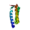
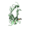
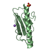
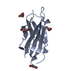
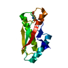
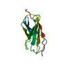
Journal: Acta Crystallogr D Biol Crystallogr / Year: 2015
Title: Combination of X-ray crystallography, SAXS and DEER to obtain the structure of the FnIII-3,4 domains of integrin α6β4.
Authors: Noelia Alonso-García / Inés García-Rubio / José A Manso / Rubén M Buey / Hector Urien / Arnoud Sonnenberg / Gunnar Jeschke / José M de Pereda /
Abstract: Integrin α6β4 is a major component of hemidesmosomes that mediate the stable anchorage of epithelial cells to the underlying basement membrane. Integrin α6β4 has also been implicated in cell ...Integrin α6β4 is a major component of hemidesmosomes that mediate the stable anchorage of epithelial cells to the underlying basement membrane. Integrin α6β4 has also been implicated in cell proliferation and migration and in carcinoma progression. The third and fourth fibronectin type III domains (FnIII-3,4) of integrin β4 mediate binding to the hemidesmosomal proteins BPAG1e and BPAG2, and participate in signalling. Here, it is demonstrated that X-ray crystallography, small-angle X-ray scattering and double electron-electron resonance (DEER) complement each other to solve the structure of the FnIII-3,4 region. The crystal structures of the individual FnIII-3 and FnIII-4 domains were solved and the relative arrangement of the FnIII domains was elucidated by combining DEER with site-directed spin labelling. Multiple structures of the interdomain linker were modelled by Monte Carlo methods complying with DEER constraints, and the final structures were selected against experimental scattering data. FnIII-3,4 has a compact and cambered flat structure with an evolutionary conserved surface that is likely to correspond to a protein-interaction site. Finally, this hybrid method is of general application for the study of other macromolecules and complexes.
History
DepositionOct 30, 2014Deposition site: RCSB / Processing site: PDBE
Revision 1.0Feb 11, 2015Provider: repository / Type: Initial release
Revision 1.1Apr 8, 2015Group: Database references
Revision 1.2May 8, 2024Group: Data collection / Database references / Category: chem_comp_atom / chem_comp_bond / database_2
Item: _database_2.pdbx_DOI / _database_2.pdbx_database_accession

-
Structure visualization

Structure viewerMolecule:
MolmilJmol/JSmol

Downloads & links

-
Assembly

Deposited unit
A: Integrin beta-4


Theoretical massNumber of molelcules
Total (without water)10,6001
Polymers10,6001
Non-polymers00
Water2,198122
1


  • Idetical with deposited unit
  • defined by author&software
TypeNameSymmetry operationNumber
identity operation1_555x,y,z1
Buried area0 Å2
ΔGint0 kcal/mol
Surface area5410 Å2
MethodPISA
Unit cell
Length a, b, c (Å)57.260, 57.260, 76.290
Angle α, β, γ (deg.)90.000, 90.000, 90.000
Int Tables number92
Space group name H-MP41212
DetailsThe biological unit is a monomer. There are 2 biological units in the asymmetric unit

-
Components

#1: Protein Integrin beta-4 / GP150


Mass: 10599.834 Da / Num. of mol.: 1 / Fragment: Fourth FnIII domain, UNP residues 1572-1666
Source method: isolated from a genetically manipulated source
Source: (gene. exp.) Homo sapiens (human) / Gene: ITGB4 / Plasmid: Modified pET15B / Production host: Escherichia coli BL21(DE3) (bacteria) / References: UniProt: P16144
#2: Water ChemComp-HOH / water


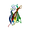
Mass: 18.015 Da / Num. of mol.: 122 / Source method: isolated from a natural source / Formula: H2O

-
Experimental details

-
Experiment

ExperimentMethod: X-RAY DIFFRACTION / Number of used crystals: 1

-
Sample preparation

CrystalDensity Matthews: 2.95 Å3/Da / Density % sol: 58.31 %
Crystal growTemperature: 277 K / Method: vapor diffusion, hanging drop / pH: 7
Details: 0.1 M Sodium Acetate (pH 4.2), 0.7 M NaH2PO4, 1.05 M K2HPO4, 2 mM DTT

-
Data collection

DiffractionMean temperature: 100 K
Diffraction sourceSource: SYNCHROTRON / Site: SLS / Beamline: X06DA / Wavelength: 0.97929 Å
DetectorType: DECTRIS PILATUS 2M-F / Detector: PIXEL / Date: Oct 21, 2013
RadiationProtocol: SINGLE WAVELENGTH / Monochromatic (M) / Laue (L): M / Scattering type: x-ray
Radiation wavelengthWavelength: 0.97929 Å / Relative weight: 1
ReflectionResolution: 1.5→45.8 Å / Num. obs: 19975 / % possible obs: 95.3 % / Observed criterion σ(I): -3 / Redundancy: 24.3 % / Biso Wilson estimate: 19.97 Å2 / Rmerge F obs: 1 / Rmerge(I) obs: 0.061 / Rrim(I) all: 0.062 / Χ2: 1.024 / Net I/σ(I): 33.88 / Num. measured all: 485023
Reflection shell

Diffraction-ID: 1 / Rejects: _

Resolution (Å)Highest resolution (Å)Rmerge F obsRmerge(I) obsMean I/σ(I) obsNum. measured obsNum. possibleNum. unique obsRrim(I) all% possible all
1.5-1.540.8881.4063.339641151215091.43399.8
1.54-1.580.9321.2133.8338442147914761.23799.8
1.58-1.630.9460.9834.6636551143214261.00399.6
1.63-1.680.9620.7136.1335786141314120.72799.9
1.68-1.730.9740.5158.0532301136713670.526100
1.73-1.790.9870.37711.4133900131813170.38599.9
1.79-1.860.990.41310.129602126411880.42194
1.86-1.940.9890.27914.091184912277340.28959.8
1.94-2.020.9980.13226.4230641118711850.13599.8
2.02-2.120.9990.10733.1130121114111410.109100
2.12-2.240.9960.11731.981574110667800.1273.2
2.24-2.370.9990.07941.042455110349890.08195.6
2.37-2.5410.0650.89223589629620.061100
2.54-2.7410.04865.37225599109100.049100
2.74-310.03879.6205688458450.038100
3-3.3510.02989.49172157647630.0399.9
3.35-3.8710.03190.73131576866140.03289.5
3.87-4.7410.022123.27142065925910.02399.8
4.74-6.7110.02121.23100574764760.021100
6.7110.019129.7557772902900.019100

-
Phasing

PhasingMethod: SIRAS

-
Processing

Software
NameVersionClassification
XDSdata reduction
XSCALEdata scaling
PHENIX1839refinement
PDB_EXTRACT3.15data extraction
SHELXDEphasing
RefinementMethod to determine structure: SIRAS / Resolution: 1.5→45.796 Å / SU ML: 0.13 / Cross valid method: FREE R-VALUE / σ(F): 1.36 / Phase error: 20.95 / Stereochemistry target values: ML
RfactorNum. reflection% reflectionSelection details
Rfree0.2181 992 4.98 %Random selection
Rwork0.1946 18911 --
obs0.1957 19903 94.93 %-
Solvent computationShrinkage radii: 0.9 Å / VDW probe radii: 1.11 Å / Solvent model: FLAT BULK SOLVENT MODEL
Displacement parametersBiso max: 109.08 Å2 / Biso mean: 28.9816 Å2 / Biso min: 11.83 Å2
Refinement stepCycle: final / Resolution: 1.5→45.796 Å
ProteinNucleic acidLigandSolventTotal
Num. atoms729 0 0 122 851
Biso mean---34.84 -
Num. residues----97
Refine LS restraints
Refine-IDTypeDev idealNumber
X-RAY DIFFRACTIONf_bond_d0.012799
X-RAY DIFFRACTIONf_angle_d1.3351089
X-RAY DIFFRACTIONf_chiral_restr0.049116
X-RAY DIFFRACTIONf_plane_restr0.005152
X-RAY DIFFRACTIONf_dihedral_angle_d13.516307
LS refinement shell

Refine-ID: X-RAY DIFFRACTION / Total num. of bins used: 7

Resolution (Å)Rfactor RfreeNum. reflection RfreeRfactor RworkNum. reflection RworkNum. reflection all% reflection obs (%)
1.5001-1.57920.25491610.233827482909100
1.5792-1.67810.23361460.218627922938100
1.6781-1.80770.25061560.207427782934100
1.8077-1.98960.26591260.24252245237180
1.9896-2.27750.26231090.20692495260488
2.2775-2.86930.20781570.197828713028100
2.8693-45.81710.19011370.17442982311998
Refinement TLS params.

Method: refined / Refine-ID: X-RAY DIFFRACTION

IDL112)L122)L132)L222)L232)L332)S11 (Å °)S12 (Å °)S13 (Å °)S21 (Å °)S22 (Å °)S23 (Å °)S31 (Å °)S32 (Å °)S33 (Å °)T112)T122)T132)T222)T232)T332)Origin x (Å)Origin y (Å)Origin z (Å)
11.705-0.66930.06920.83080.87461.52490.0045-0.1826-0.09830.3136-0.03880.01740.19190.03910.00070.17770.00060.00330.1560.00180.12567.768424.035916.1109
21.6990.72230.20230.47950.58221.5823-0.037-0.0322-0.30690.3019-0.04830.23390.0724-0.1546-0.00140.1893-0.00510.04170.12920.01560.18325.531414.338710.8475
33.99993.8122-2.52893.8611-1.69943.77770.37030.3971.0402-0.3961-0.01250.5948-0.6309-1.20420.37560.20280.0524-0.04580.3541-0.01720.2699-4.157531.74954.9183
41.1710.48510.00580.9264-0.08350.69320.0021-0.03060.09340.0252-0.0012-0.32410.05680.0742-0.00030.21220.0156-0.01140.1916-0.00850.13749.367324.84669.3999
53.5043-0.2404-0.17821.3161-0.84691.69320.15630.1595-0.22-0.1353-0.27240.20190.0762-0.1496-0.00560.1546-0.0096-0.00640.17090.00560.13152.25720.862610.8149
Refinement TLS group
IDRefine-IDRefine TLS-IDSelection detailsAuth asym-IDAuth seq-ID
1X-RAY DIFFRACTION1chain 'A' and (resid 1570 through 1592 )A0
2X-RAY DIFFRACTION2chain 'A' and (resid 1593 through 1610 )A0
3X-RAY DIFFRACTION3chain 'A' and (resid 1611 through 1617 )A0
4X-RAY DIFFRACTION4chain 'A' and (resid 1618 through 1641 )A0
5X-RAY DIFFRACTION5chain 'A' and (resid 1642 through 1666 )A0

+
About Yorodumi

-
News

-
Feb 9, 2022. New format data for meta-information of EMDB entries

New format data for meta-information of EMDB entries

  • Version 3 of the EMDB header file is now the official format.
  • The previous official version 1.9 will be removed from the archive.

Related info.:EMDB header

External links:wwPDB to switch to version 3 of the EMDB data model

-
Aug 12, 2020. Covid-19 info

Covid-19 info

URL: https://pdbjlc1.pdbj.org/emnavi/covid19.php

New page: Covid-19 featured information page in EM Navigator.

Related info.:Covid-19 info / Mar 5, 2020. Novel coronavirus structure data

+
Mar 5, 2020. Novel coronavirus structure data

Novel coronavirus structure data

Related info.:Yorodumi Speices / Aug 12, 2020. Covid-19 info

External links:COVID-19 featured content - PDBj / Molecule of the Month (242):Coronavirus Proteases

+
Jan 31, 2019. EMDB accession codes are about to change! (news from PDBe EMDB page)

EMDB accession codes are about to change! (news from PDBe EMDB page)

  • The allocation of 4 digits for EMDB accession codes will soon come to an end. Whilst these codes will remain in use, new EMDB accession codes will include an additional digit and will expand incrementally as the available range of codes is exhausted. The current 4-digit format prefixed with “EMD-” (i.e. EMD-XXXX) will advance to a 5-digit format (i.e. EMD-XXXXX), and so on. It is currently estimated that the 4-digit codes will be depleted around Spring 2019, at which point the 5-digit format will come into force.
  • The EM Navigator/Yorodumi systems omit the EMD- prefix.

Related info.:Q: What is EMD? / ID/Accession-code notation in Yorodumi/EM Navigator

External links:EMDB Accession Codes are Changing Soon! / Contact to PDBj

+
Jul 12, 2017. Major update of PDB

Major update of PDB

  • wwPDB released updated PDB data conforming to the new PDBx/mmCIF dictionary.
  • This is a major update changing the version number from 4 to 5, and with Remediation, in which all the entries are updated.
  • In this update, many items about electron microscopy experimental information are reorganized (e.g. em_software).
  • Now, EM Navigator and Yorodumi are based on the updated data.

External links:wwPDB Remediation / Enriched Model Files Conforming to OneDep Data Standards Now Available in the PDB FTP Archive

-
Yorodumi

Thousand views of thousand structures

  • Yorodumi is a browser for structure data from EMDB, PDB, SASBDB, etc.
  • This page is also the successor to EM Navigator detail page, and also detail information page/front-end page for Omokage search.
  • The word "yorodu" (or yorozu) is an old Japanese word meaning "ten thousand". "mi" (miru) is to see.

Related info.:EMDB / PDB / SASBDB / Comparison of 3 databanks / Yorodumi Search / Aug 31, 2016. New EM Navigator & Yorodumi / Yorodumi Papers / Jmol/JSmol / Function and homology information / Changes in new EM Navigator and Yorodumi

Read more