 Movie
Movie Controller

[English] 日本語
 Yorodumi
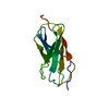
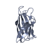
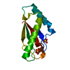
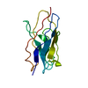
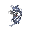
- PDB-4g4v: Crystal structure of peptidoglycan-associated lipoprotein from Ac... -
+ Open data

- Basic information
| Entry | Database: PDB / ID: 4g4v | |||||||||
|---|---|---|---|---|---|---|---|---|---|---|
| Title | Crystal structure of peptidoglycan-associated lipoprotein from Acinetobacter baumannii | |||||||||
 Components Components | Peptidoglycan-associated lipoprotein | |||||||||
 Keywords Keywords | PEPTIDE BINDING PROTEIN / OmpA-like domain / MEMBRANE PROTEIN | |||||||||
| Function / homology | OmpA-like domain / 60s Ribosomal Protein L30; Chain: A; / 2-Layer Sandwich / Alpha Beta / 2,6-DIAMINOPIMELIC ACID / Peptidoglycan-associated lipoprotein / Peptidoglycan-associated lipoprotein Function and homology information Function and homology information | |||||||||
| Biological species |  Acinetobacter baumannii (bacteria) Acinetobacter baumannii (bacteria) | |||||||||
| Method |  X-RAY DIFFRACTION / SYNCHROTRON / MOLECULAR REPLACEMENT / Resolution: 1.9 Å X-RAY DIFFRACTION / SYNCHROTRON / MOLECULAR REPLACEMENT / Resolution: 1.9 Å | |||||||||
 Authors Authors | Lee, W.C. / Song, J.H. / Park, J.S. / Kim, H.Y. | |||||||||
 Citation Citation | Journal: To be Published Title: Enantiomer-dependent amino acid binding affinity of OmpA-like domains from Acinetobacter baumannii peptidoglycan-associated lipoprotein and OmpA Authors: Lee, W.C. / Park, J.S. / Song, J.H. / Kim, S.I. / Lee, J.C. / Cheong, J. / Kim, H.Y. | |||||||||
| History |
|
- Structure visualization
| Structure viewer | Molecule: MolmilJmol/JSmol |
|---|
- Downloads & links
-Download
| PDBx/mmCIF format | 4g4v.cif.gz | 37.1 KB | Display | PDBx/mmCIF format |
|---|---|---|---|---|
| PDB format | pdb4g4v.ent.gz | 23.7 KB | Display | PDB format |
| PDBx/mmJSON format | 4g4v.json.gz | Tree view | PDBx/mmJSON format | |
| Others |  Other downloads Other downloads |
-Validation report
| Arichive directory | https://data.pdbj.org/pub/pdb/validation_reports/g4/4g4vftp://data.pdbj.org/pub/pdb/validation_reports/g4/4g4v | HTTPS FTP |
|---|
-Related structure data
| Related structure data |  4g4wC 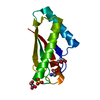 4g4xC  4g4yC  4g4zC  4g88C  3td4S C: citing same article ( S: Starting model for refinement |
|---|---|
| Similar structure data |


-Links
 PDBj
PDBj- Assembly
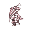
| Deposited unit | 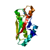
| ||||||||
|---|---|---|---|---|---|---|---|---|---|
| 1 |
| ||||||||
| Unit cell |
|
-Components
| #1: Protein | Mass: 12814.040 Da / Num. of mol.: 1 / Fragment: UNP residues 75-184 Source method: isolated from a genetically manipulated source Source: (gene. exp.) Acinetobacter baumannii (bacteria) / Strain: TCDC-AB0715 / Gene: pal / Plasmid: pET28a / Production host: |
|---|---|
| #2: Chemical | ChemComp-API / 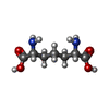  Type: L-peptide linking / Mass: 190.197 Da / Num. of mol.: 1 / Source method: obtained synthetically / Formula: C7H14N2O4 Type: L-peptide linking / Mass: 190.197 Da / Num. of mol.: 1 / Source method: obtained synthetically / Formula: C7H14N2O4 |
| #3: Water | ChemComp-HOH /  Mass: 18.015 Da / Num. of mol.: 50 / Source method: isolated from a natural source / Formula: H2O Mass: 18.015 Da / Num. of mol.: 50 / Source method: isolated from a natural source / Formula: H2O |
-Experimental details
-Experiment
| Experiment | Method: X-RAY DIFFRACTION / Number of used crystals: 1 |
|---|
- Sample preparation
| Crystal | Density Matthews: 1.85 Å3/Da / Density % sol: 33.63 % / Mosaicity: 1.278 ° |
|---|---|
| Crystal grow | Temperature: 293 K / Method: vapor diffusion / pH: 4.2 Details: 0.1M PHOSPHATE CITRATE, 30% PEG 3350, pH 4.2, VAPOR DIFFUSION, temperature 293K |
-Data collection
| Diffraction | Mean temperature: 100 K | |||||||||||||||||||||||||||||||||||||||||||||||||||||||||||||||||||||||||||||
|---|---|---|---|---|---|---|---|---|---|---|---|---|---|---|---|---|---|---|---|---|---|---|---|---|---|---|---|---|---|---|---|---|---|---|---|---|---|---|---|---|---|---|---|---|---|---|---|---|---|---|---|---|---|---|---|---|---|---|---|---|---|---|---|---|---|---|---|---|---|---|---|---|---|---|---|---|---|---|
| Diffraction source | Source: SYNCHROTRON / Site: Photon Factory  / Beamline: BL-17A / Wavelength: 1 Å / Beamline: BL-17A / Wavelength: 1 Å | |||||||||||||||||||||||||||||||||||||||||||||||||||||||||||||||||||||||||||||
| Detector | Type: ADSC QUANTUM 270 / Detector: CCD / Date: Jul 3, 2011 | |||||||||||||||||||||||||||||||||||||||||||||||||||||||||||||||||||||||||||||
| Radiation | Protocol: SINGLE WAVELENGTH / Monochromatic (M) / Laue (L): M / Scattering type: x-ray | |||||||||||||||||||||||||||||||||||||||||||||||||||||||||||||||||||||||||||||
| Radiation wavelength | Wavelength: 1 Å / Relative weight: 1 | |||||||||||||||||||||||||||||||||||||||||||||||||||||||||||||||||||||||||||||
| Reflection | Resolution: 1.9→50 Å / Num. obs: 7884 / % possible obs: 99.7 % / Redundancy: 6.5 % / Rmerge(I) obs: 0.071 / Χ2: 3.123 / Net I/σ(I): 16.9 | |||||||||||||||||||||||||||||||||||||||||||||||||||||||||||||||||||||||||||||
| Reflection shell |
|
- Processing
| Software |
| |||||||||||||||||||||||||||||||||||||||||||||||||||||||||||||||||||||||||||||
|---|---|---|---|---|---|---|---|---|---|---|---|---|---|---|---|---|---|---|---|---|---|---|---|---|---|---|---|---|---|---|---|---|---|---|---|---|---|---|---|---|---|---|---|---|---|---|---|---|---|---|---|---|---|---|---|---|---|---|---|---|---|---|---|---|---|---|---|---|---|---|---|---|---|---|---|---|---|---|
| Refinement | Method to determine structure: MOLECULAR REPLACEMENT Starting model: PDB ENTRY 3TD4 Resolution: 1.9→50 Å / Occupancy max: 1 / Occupancy min: 1 / σ(F): 0
| |||||||||||||||||||||||||||||||||||||||||||||||||||||||||||||||||||||||||||||
| Solvent computation | Bsol: 39.427 Å2 | |||||||||||||||||||||||||||||||||||||||||||||||||||||||||||||||||||||||||||||
| Displacement parameters | Biso max: 62.46 Å2 / Biso mean: 33.1362 Å2 / Biso min: 17.66 Å2 | |||||||||||||||||||||||||||||||||||||||||||||||||||||||||||||||||||||||||||||
| Refinement step | Cycle: LAST / Resolution: 1.9→50 Å
| |||||||||||||||||||||||||||||||||||||||||||||||||||||||||||||||||||||||||||||
| Refine LS restraints |
| |||||||||||||||||||||||||||||||||||||||||||||||||||||||||||||||||||||||||||||
| LS refinement shell | Refine-ID: X-RAY DIFFRACTION / Total num. of bins used: 10
| |||||||||||||||||||||||||||||||||||||||||||||||||||||||||||||||||||||||||||||
| Xplor file |
|
