Movie
Movie Controller

[English] 日本語
 Yorodumi
- EMDB-6475: Cryo-EM structure of the rabbit voltage-gated calcium channel Cav... -
+ Open data

- Basic information
| Entry | Database: EMDB / ID: EMD-6475 | |||||||||
|---|---|---|---|---|---|---|---|---|---|---|
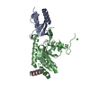
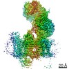
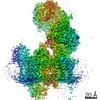
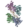
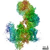
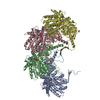
| Title | Cryo-EM structure of the rabbit voltage-gated calcium channel Cav1.1 at 4.2 angstrom resolution | |||||||||
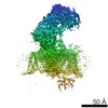
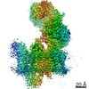
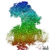
 Map data Map data | Reconstruction of a membrane protein | |||||||||
 Sample Sample |
| |||||||||
| Function / homology |  Function and homology information Function and homology informationvoltage-gated calcium channel activity involved in regulation of presynaptic cytosolic calcium levels / Phase 0 - rapid depolarisation / Phase 2 - plateau phase / voltage-gated calcium channel activity involved in AV node cell action potential / positive regulation of calcium ion transmembrane transport via high voltage-gated calcium channel / Regulation of insulin secretion / Presynaptic depolarization and calcium channel opening / membrane depolarization during atrial cardiac muscle cell action potential / photoreceptor ribbon synapse / high voltage-gated calcium channel activity ...voltage-gated calcium channel activity involved in regulation of presynaptic cytosolic calcium levels / Phase 0 - rapid depolarisation / Phase 2 - plateau phase / voltage-gated calcium channel activity involved in AV node cell action potential / positive regulation of calcium ion transmembrane transport via high voltage-gated calcium channel / Regulation of insulin secretion / Presynaptic depolarization and calcium channel opening / membrane depolarization during atrial cardiac muscle cell action potential / photoreceptor ribbon synapse / high voltage-gated calcium channel activity / membrane depolarization during AV node cell action potential / L-type voltage-gated calcium channel complex / positive regulation of muscle contraction / calcium ion import / positive regulation of calcium ion transport / regulation of release of sequestered calcium ion into cytosol / regulation of calcium ion transmembrane transport via high voltage-gated calcium channel / cellular response to caffeine / voltage-gated calcium channel complex / regulation of heart rate by cardiac conduction / calcium ion import across plasma membrane / voltage-gated calcium channel activity / release of sequestered calcium ion into cytosol / visual perception / T-tubule / muscle contraction / protein localization to plasma membrane / calcium channel regulator activity / phosphoprotein binding / sarcolemma / calcium ion transmembrane transport / actin filament binding / calcium ion transport / presynapse / chemical synaptic transmission / transmembrane transporter binding / calmodulin binding / protein domain specific binding / protein kinase binding / metal ion binding / identical protein binding / plasma membrane Similarity search - Function | |||||||||
| Biological species |  | |||||||||
| Method | single particle reconstruction / cryo EM / Resolution: 4.2 Å | |||||||||
 Authors Authors | Wu JP / Yan Z / Li ZQ / Yan N | |||||||||
 Citation Citation | Journal: Science / Year: 2015 Title: Structure of the voltage-gated calcium channel Cav1.1 complex. Authors: Jianping Wu / Zhen Yan / Zhangqiang Li / Chuangye Yan / Shan Lu / Mengqiu Dong / Nieng Yan /  Abstract: The voltage-gated calcium channel Ca(v)1.1 is engaged in the excitation-contraction coupling of skeletal muscles. The Ca(v)1.1 complex consists of the pore-forming subunit α1 and auxiliary subunits ...The voltage-gated calcium channel Ca(v)1.1 is engaged in the excitation-contraction coupling of skeletal muscles. The Ca(v)1.1 complex consists of the pore-forming subunit α1 and auxiliary subunits α2δ, β, and γ. We report the structure of the rabbit Ca(v)1.1 complex determined by single-particle cryo-electron microscopy. The four homologous repeats of the α1 subunit are arranged clockwise in the extracellular view. The γ subunit, whose structure resembles claudins, interacts with the voltage-sensing domain of repeat IV (VSD(IV)), whereas the cytosolic β subunit is located adjacent to VSD(II) of α1. The α2 subunit interacts with the extracellular loops of repeats I to III through its VWA and Cache1 domains. The structure reveals the architecture of a prototypical eukaryotic Ca(v) channel and provides a framework for understanding the function and disease mechanisms of Ca(v) and Na(v) channels. | |||||||||
| History |
|
- Structure visualization
| Movie |
Movie viewer |
|---|---|
| Structure viewer | EM map: SurfViewMolmilJmol/JSmol |
| Supplemental images |

- Downloads & links
-EMDB archive
| Map data | emd_6475.map.gz | 59.7 MB | EMDB map data format | |
|---|---|---|---|---|
| Header (meta data) | emd-6475-v30.xmlemd-6475.xml | 12.2 KB 12.2 KB | Display Display | EMDB header |
| Images |  400_6475.gif 400_6475.gif 80_6475.gif 80_6475.gif | 49.8 KB 3.4 KB | ||
| Archive directory |  http://ftp.pdbj.org/pub/emdb/structures/EMD-6475ftp://ftp.pdbj.org/pub/emdb/structures/EMD-6475 http://ftp.pdbj.org/pub/emdb/structures/EMD-6475ftp://ftp.pdbj.org/pub/emdb/structures/EMD-6475 | HTTPS FTP |
-Related structure data
| Related structure data |  3jbrMC 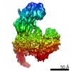 6476C M: atomic model generated by this map C: citing same article ( |
|---|---|
| Similar structure data |


-Links
| EMDB pages | EMDB (EBI/PDBe) / EMDataResource |
|---|---|
| Related items in Molecule of the Month |


-Map
| File | Download / File: emd_6475.map.gz / Format: CCP4 / Size: 62.5 MB / Type: IMAGE STORED AS FLOATING POINT NUMBER (4 BYTES) | ||||||||||||||||||||||||||||||||||||||||||||||||||||||||||||||||||||
|---|---|---|---|---|---|---|---|---|---|---|---|---|---|---|---|---|---|---|---|---|---|---|---|---|---|---|---|---|---|---|---|---|---|---|---|---|---|---|---|---|---|---|---|---|---|---|---|---|---|---|---|---|---|---|---|---|---|---|---|---|---|---|---|---|---|---|---|---|---|
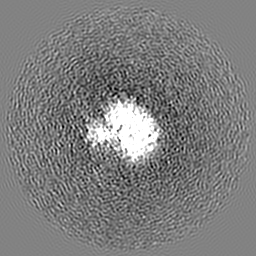
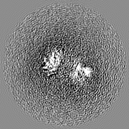
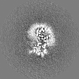
| Annotation | Reconstruction of a membrane protein | ||||||||||||||||||||||||||||||||||||||||||||||||||||||||||||||||||||
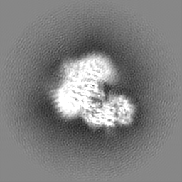
| Projections & slices | Image control
Images are generated by Spider. | ||||||||||||||||||||||||||||||||||||||||||||||||||||||||||||||||||||
| Voxel size | X=Y=Z: 1.32 Å | ||||||||||||||||||||||||||||||||||||||||||||||||||||||||||||||||||||
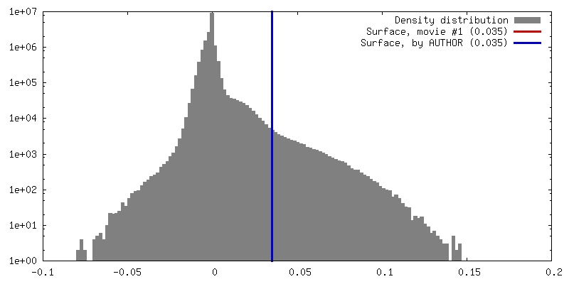
| Density |
| ||||||||||||||||||||||||||||||||||||||||||||||||||||||||||||||||||||
| Symmetry | Space group: 1 | ||||||||||||||||||||||||||||||||||||||||||||||||||||||||||||||||||||
| Details | EMDB XML:
CCP4 map header:
| ||||||||||||||||||||||||||||||||||||||||||||||||||||||||||||||||||||
 Z (Sec.)
 Y (Row.)
 X (Col.)


-Supplemental data
- Sample components
-Entire : a membrane protein cryo-sample
| Entire | Name: a membrane protein cryo-sample |
|---|---|
| Components |
|
-Supramolecule #1000: a membrane protein cryo-sample
| Supramolecule | Name: a membrane protein cryo-sample / type: sample / ID: 1000 / Details: The sample was monodisperse / Number unique components: 4 |
|---|---|
| Molecular weight | Theoretical: 450 KDa |
-Macromolecule #1: Voltage-dependent L-type calcium channel subunit alpha-1S
| Macromolecule | Name: Voltage-dependent L-type calcium channel subunit alpha-1S type: protein_or_peptide / ID: 1 / Number of copies: 1 / Oligomeric state: monomer / Recombinant expression: No / Database: NCBI |
|---|---|
| Source (natural) | Organism: |
| Molecular weight | Theoretical: 210 KDa |
| Sequence | UniProtKB: Voltage-dependent L-type calcium channel subunit alpha-1S |
-Macromolecule #2: Voltage-dependent L-type calcium channel subunit beta-2
| Macromolecule | Name: Voltage-dependent L-type calcium channel subunit beta-2 type: protein_or_peptide / ID: 2 / Number of copies: 1 / Oligomeric state: monomer / Recombinant expression: No / Database: NCBI |
|---|---|
| Source (natural) | Organism: |
| Molecular weight | Theoretical: 30 KDa |
| Sequence | UniProtKB: Voltage-dependent L-type calcium channel subunit beta-2 |
-Macromolecule #3: Voltage-dependent calcium channel gamma-1 subunit
| Macromolecule | Name: Voltage-dependent calcium channel gamma-1 subunit / type: protein_or_peptide / ID: 3 / Number of copies: 1 / Oligomeric state: monomer / Recombinant expression: No / Database: NCBI |
|---|---|
| Source (natural) | Organism: |
| Molecular weight | Theoretical: 25 KDa |
| Sequence | UniProtKB: Voltage-dependent calcium channel gamma-1 subunit |
-Macromolecule #4: Voltage-dependent calcium channel subunit alpha-2/delta-1
| Macromolecule | Name: Voltage-dependent calcium channel subunit alpha-2/delta-1 type: protein_or_peptide / ID: 4 / Number of copies: 1 / Oligomeric state: monomer / Recombinant expression: No / Database: NCBI |
|---|---|
| Source (natural) | Organism: |
| Molecular weight | Theoretical: 125 KDa |
| Sequence | UniProtKB: Voltage-dependent calcium channel subunit alpha-2/delta-1 |
-Experimental details
-Structure determination
| Method | cryo EM |
|---|---|
 Processing Processing | single particle reconstruction |
| Aggregation state | particle |
-Sample preparation
| Concentration | 0.1 mg/mL |
|---|---|
| Buffer | pH: 7.4 / Details: 200mM NaCl, 20mM MOPS, 0.5mM CaCl2, 0.1% digitonin |
| Grid | Details: carbon coated grid |
| Vitrification | Cryogen name: ETHANE / Chamber humidity: 100 % / Instrument: FEI VITROBOT MARK IV / Method: Blot for 2 seconds before plunging |
- Electron microscopy
| Microscope | FEI TITAN KRIOS |
|---|---|
| Date | Feb 2, 2015 |
| Image recording | Category: CCD / Film or detector model: GATAN K2 (4k x 4k) / Number real images: 3991 / Average electron dose: 50 e/Å2 |
| Electron beam | Acceleration voltage: 300 kV / Electron source:  FIELD EMISSION GUN FIELD EMISSION GUN |
| Electron optics | Illumination mode: SPOT SCAN / Imaging mode: BRIGHT FIELD / Cs: 2.7 mm / Nominal defocus max: 0.0033 µm / Nominal defocus min: 0.002 µm |
| Sample stage | Specimen holder model: FEI TITAN KRIOS AUTOGRID HOLDER |
| Experimental equipment |  Model: Titan Krios / Image courtesy: FEI Company |
-Image processing
| Details | The particles were selected using RELION. |
|---|---|
| CTF correction | Details: each micrograph |
| Final reconstruction | Resolution.type: BY AUTHOR / Resolution: 4.2 Å / Resolution method: OTHER / Software - Name: RELION / Number images used: 353372 |