 Movie
Movie Controller

[English] 日本語
 Yorodumi
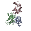
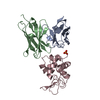
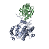
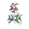
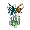
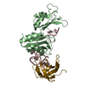
- PDB-1t0j: Crystal structure of a complex between voltage-gated calcium chan... -
+ Open data

- Basic information
| Entry | Database: PDB / ID: 1t0j | ||||||
|---|---|---|---|---|---|---|---|
| Title | Crystal structure of a complex between voltage-gated calcium channel beta2a subunit and a peptide of the alpha1c subunit | ||||||
 Components Components |
| ||||||
 Keywords Keywords | SIGNALING PROTEIN / SH3 domain / nucleotide kinase like domain / ion channel / calcium channel / AID | ||||||
| Function / homology |  Function and homology information Function and homology informationvoltage-gated calcium channel activity involved in regulation of presynaptic cytosolic calcium levels / Phase 0 - rapid depolarisation / Phase 2 - plateau phase / voltage-gated calcium channel activity involved in AV node cell action potential / voltage-gated calcium channel activity involved in cardiac muscle cell action potential / positive regulation of calcium ion transmembrane transport via high voltage-gated calcium channel / Regulation of insulin secretion / immune system development / positive regulation of adenylate cyclase activity / Presynaptic depolarization and calcium channel opening ...voltage-gated calcium channel activity involved in regulation of presynaptic cytosolic calcium levels / Phase 0 - rapid depolarisation / Phase 2 - plateau phase / voltage-gated calcium channel activity involved in AV node cell action potential / voltage-gated calcium channel activity involved in cardiac muscle cell action potential / positive regulation of calcium ion transmembrane transport via high voltage-gated calcium channel / Regulation of insulin secretion / immune system development / positive regulation of adenylate cyclase activity / Presynaptic depolarization and calcium channel opening / membrane depolarization during atrial cardiac muscle cell action potential / calcium ion transmembrane transport via high voltage-gated calcium channel / Phase 2 - plateau phase / photoreceptor ribbon synapse / high voltage-gated calcium channel activity / cardiac conduction / embryonic forelimb morphogenesis / membrane depolarization during AV node cell action potential / L-type voltage-gated calcium channel complex / membrane depolarization during cardiac muscle cell action potential / cell communication by electrical coupling involved in cardiac conduction / positive regulation of muscle contraction / calcium ion import / NCAM1 interactions / camera-type eye development / regulation of ventricular cardiac muscle cell action potential / positive regulation of calcium ion transport / cardiac muscle cell action potential involved in contraction / calcium ion transport into cytosol / voltage-gated calcium channel complex / Phase 0 - rapid depolarisation / regulation of heart rate by cardiac conduction / alpha-actinin binding / calcium ion import across plasma membrane / voltage-gated calcium channel activity / regulation of cardiac muscle contraction by regulation of the release of sequestered calcium ion / visual perception / protein localization to plasma membrane / calcium channel regulator activity / Regulation of insulin secretion / phosphoprotein binding / postsynaptic density membrane / Z disc / calcium ion transmembrane transport / calcium ion transport / actin filament binding / Adrenaline,noradrenaline inhibits insulin secretion / heart development / positive regulation of cytosolic calcium ion concentration / presynapse / chemical synaptic transmission / perikaryon / calmodulin binding / postsynaptic density / protein domain specific binding / dendrite / protein kinase binding / membrane / metal ion binding / identical protein binding / plasma membrane / cytoplasm Similarity search - Function | ||||||
| Biological species |   Homo sapiens (human) Homo sapiens (human) | ||||||
| Method |  X-RAY DIFFRACTION / SYNCHROTRON / MOLECULAR REPLACEMENT / Resolution: 2 Å X-RAY DIFFRACTION / SYNCHROTRON / MOLECULAR REPLACEMENT / Resolution: 2 Å | ||||||
 Authors Authors | Van Petegem, F. / Clark, K. / Chatelain, F. / Minor Jr., D. | ||||||
 Citation Citation | Journal: Nature / Year: 2004 Title: Structure of a complex between a voltage-gated calcium channel beta-subunit and an alpha-subunit domain. Authors: Van Petegem, F. / Clark, K.A. / Chatelain, F.C. / Minor, D.L. | ||||||
| History |
|
- Structure visualization
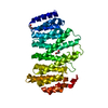
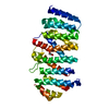
| Structure viewer | Molecule: MolmilJmol/JSmol |
|---|
- Downloads & links
-Download
| PDBx/mmCIF format | 1t0j.cif.gz | 78 KB | Display | PDBx/mmCIF format |
|---|---|---|---|---|
| PDB format | pdb1t0j.ent.gz | 56.2 KB | Display | PDB format |
| PDBx/mmJSON format | 1t0j.json.gz | Tree view | PDBx/mmJSON format | |
| Others |  Other downloads Other downloads |
-Validation report
| Arichive directory | https://data.pdbj.org/pub/pdb/validation_reports/t0/1t0jftp://data.pdbj.org/pub/pdb/validation_reports/t0/1t0j | HTTPS FTP |
|---|
-Related structure data
| Related structure data |  1t0hC  1tohS S: Starting model for refinement C: citing same article ( |
|---|---|
| Similar structure data |


-Links
 PDBj
PDBj


- Assembly
| Deposited unit | 
| ||||||||
|---|---|---|---|---|---|---|---|---|---|
| 1 |
| ||||||||
| 2 | 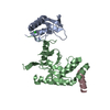
| ||||||||
| Unit cell |
|
-Components
| #1: Protein | Mass: 14996.776 Da / Num. of mol.: 1 / Fragment: residues 17-145 Source method: isolated from a genetically manipulated source Source: (gene. exp.)  |
|---|---|
| #2: Protein | Mass: 25321.291 Da / Num. of mol.: 1 / Fragment: residues 203-425 Source method: isolated from a genetically manipulated source Source: (gene. exp.) |
| #3: Protein/peptide | Mass: 2308.476 Da / Num. of mol.: 1 / Fragment: Residues 428-443 Source method: isolated from a genetically manipulated source Source: (gene. exp.) Homo sapiens (human) / Gene: CACNA1C, CACNL1A1, CCHL1A1, CACH2, CACN2 / Plasmid: modified pET27 / Production host: |
| #4: Chemical | ChemComp-CL /   Mass: 35.453 Da / Num. of mol.: 1 / Source method: obtained synthetically / Formula: Cl Mass: 35.453 Da / Num. of mol.: 1 / Source method: obtained synthetically / Formula: Cl |
| #5: Water | ChemComp-HOH /  Mass: 18.015 Da / Num. of mol.: 138 / Source method: isolated from a natural source / Formula: H2O Mass: 18.015 Da / Num. of mol.: 138 / Source method: isolated from a natural source / Formula: H2O |
-Experimental details
-Experiment
| Experiment | Method: X-RAY DIFFRACTION / Number of used crystals: 1 |
|---|
- Sample preparation
| Crystal | Density Matthews: 2.4 Å3/Da / Density % sol: 48.8 % |
|---|---|
| Crystal grow | Temperature: 277 K / Method: vapor diffusion, hanging drop / pH: 8 Details: Tris-Cl, PEG 4000, NaCl, pH 8, VAPOR DIFFUSION, HANGING DROP, temperature 277K |
-Data collection
| Diffraction | Mean temperature: 100 K |
|---|---|
| Diffraction source | Source: SYNCHROTRON / Site: ALS  / Beamline: 8.3.1 / Wavelength: 1.1159 Å / Beamline: 8.3.1 / Wavelength: 1.1159 Å |
| Detector | Type: ADSC QUANTUM 210 / Detector: CCD / Date: Feb 7, 2004 |
| Radiation | Monochromator: Double crystal / Protocol: SINGLE WAVELENGTH / Monochromatic (M) / Laue (L): M / Scattering type: x-ray |
| Radiation wavelength | Wavelength: 1.1159 Å / Relative weight: 1 |
| Reflection | Resolution: 2→30 Å / Num. all: 24890 / Num. obs: 24890 / % possible obs: 97.3 % / Observed criterion σ(F): 0 / Observed criterion σ(I): 1 / Redundancy: 2.49 % / Biso Wilson estimate: 21.503 Å2 / Rsym value: 0.081 / Net I/σ(I): 11.65 |
| Reflection shell | Resolution: 2→2.07 Å / Redundancy: 2.54 % / Mean I/σ(I) obs: 2.06 / Num. unique all: 2503 / Rsym value: 0.385 / % possible all: 96.1 |
- Processing
| Software |
| ||||||||||||||||||||||||||||||||||||||||||||||||||||||||||||||||||||||||||||||||||||||||||||||||||||||||||||||||||||||||||||||||||||||||||||||||||||||||||||||||
|---|---|---|---|---|---|---|---|---|---|---|---|---|---|---|---|---|---|---|---|---|---|---|---|---|---|---|---|---|---|---|---|---|---|---|---|---|---|---|---|---|---|---|---|---|---|---|---|---|---|---|---|---|---|---|---|---|---|---|---|---|---|---|---|---|---|---|---|---|---|---|---|---|---|---|---|---|---|---|---|---|---|---|---|---|---|---|---|---|---|---|---|---|---|---|---|---|---|---|---|---|---|---|---|---|---|---|---|---|---|---|---|---|---|---|---|---|---|---|---|---|---|---|---|---|---|---|---|---|---|---|---|---|---|---|---|---|---|---|---|---|---|---|---|---|---|---|---|---|---|---|---|---|---|---|---|---|---|---|---|---|---|
| Refinement | Method to determine structure: MOLECULAR REPLACEMENT Starting model: PDB ENTRY 1TOH Resolution: 2→30 Å / Cor.coef. Fo:Fc: 0.938 / Cor.coef. Fo:Fc free: 0.916 / SU B: 4.124 / SU ML: 0.115 / TLS residual ADP flag: LIKELY RESIDUAL / Cross valid method: THROUGHOUT / σ(F): 0 / σ(I): 0 / ESU R: 0.178 / ESU R Free: 0.163 / Stereochemistry target values: MAXIMUM LIKELIHOOD
| ||||||||||||||||||||||||||||||||||||||||||||||||||||||||||||||||||||||||||||||||||||||||||||||||||||||||||||||||||||||||||||||||||||||||||||||||||||||||||||||||
| Solvent computation | Ion probe radii: 0.8 Å / Shrinkage radii: 0.8 Å / VDW probe radii: 1.4 Å / Solvent model: BABINET MODEL WITH MASK | ||||||||||||||||||||||||||||||||||||||||||||||||||||||||||||||||||||||||||||||||||||||||||||||||||||||||||||||||||||||||||||||||||||||||||||||||||||||||||||||||
| Displacement parameters | Biso mean: 20.58 Å2
| ||||||||||||||||||||||||||||||||||||||||||||||||||||||||||||||||||||||||||||||||||||||||||||||||||||||||||||||||||||||||||||||||||||||||||||||||||||||||||||||||
| Refinement step | Cycle: LAST / Resolution: 2→30 Å
| ||||||||||||||||||||||||||||||||||||||||||||||||||||||||||||||||||||||||||||||||||||||||||||||||||||||||||||||||||||||||||||||||||||||||||||||||||||||||||||||||
| Refine LS restraints |
| ||||||||||||||||||||||||||||||||||||||||||||||||||||||||||||||||||||||||||||||||||||||||||||||||||||||||||||||||||||||||||||||||||||||||||||||||||||||||||||||||
| LS refinement shell | Resolution: 2→2.052 Å / Total num. of bins used: 20 /
| ||||||||||||||||||||||||||||||||||||||||||||||||||||||||||||||||||||||||||||||||||||||||||||||||||||||||||||||||||||||||||||||||||||||||||||||||||||||||||||||||
| Refinement TLS params. | Method: refined / Refine-ID: X-RAY DIFFRACTION
| ||||||||||||||||||||||||||||||||||||||||||||||||||||||||||||||||||||||||||||||||||||||||||||||||||||||||||||||||||||||||||||||||||||||||||||||||||||||||||||||||
| Refinement TLS group |
|
