Movie
Movie Controller


+ Open data

- Basic information
| Entry |  | ||||||||||||
|---|---|---|---|---|---|---|---|---|---|---|---|---|---|
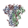
| Title | Cryo-EM structure of GD-BatCoV (BtCoV/Ii/GD/2014-422) S-trimer | ||||||||||||
 Map data Map data | |||||||||||||
 Sample Sample |
| ||||||||||||
 Keywords Keywords | spike protein / VIRAL PROTEIN | ||||||||||||
| Function / homology |  Function and homology information Function and homology informationvirion component / host cell endoplasmic reticulum-Golgi intermediate compartment membrane / receptor-mediated virion attachment to host cell / endocytosis involved in viral entry into host cell / fusion of virus membrane with host plasma membrane / fusion of virus membrane with host endosome membrane / viral envelope / host cell plasma membrane / virion membrane / membrane Similarity search - Function | ||||||||||||
| Biological species |   Middle East respiratory syndrome-related coronavirus / Tequatrovirus T4 Middle East respiratory syndrome-related coronavirus / Tequatrovirus T4 | ||||||||||||
| Method | single particle reconstruction / cryo EM / Resolution: 3.1 Å | ||||||||||||
 Authors Authors | Yuan H / Xiong X / Gao X / Li Z / Wang J | ||||||||||||
| Funding support | 3 items
| ||||||||||||
 Citation Citation | Journal: Sci Adv / Year: 2025 Title: Structures and receptor binding activities of merbecovirus spike proteins reveal key signatures for human DPP4 adaptation. Authors: Hang Yuan / Jingjing Wang / Yong Ma / Zimu Li / Xijie Gao / Gul Habib / Banghui Liu / Jing Chen / Jun He / Peng Zhou / Zheng-Li Shi / Xinwen Chen / Xiaoli Xiong /  Abstract: Merbecoviruses from bats, pangolins, and hedgehogs pose significant zoonotic threats, with a limited understanding of receptor binding by their spike (S) proteins. Here, we report cryo-EM structures ...Merbecoviruses from bats, pangolins, and hedgehogs pose significant zoonotic threats, with a limited understanding of receptor binding by their spike (S) proteins. Here, we report cryo-EM structures of GD-BatCoV (BtCoV-422) and SE-PangolinCoV (MjHKU4r-CoV-1) RBDs in complex with human DPP4 (hDPP4). These structures exhibit a substantial offset in their hDPP4 interaction interfaces, revealing a conserved hydrophobic cluster as a convergent signature of DPP4 binding within the MERS-HKU4 clade of merbecoviruses. Structure-guided mutagenesis demonstrates that favorable interactions are distributed across multiple receptor binding motif (RBM) regions, working synergistically to confer high-affinity hDPP4 binding. Swapping of the merbecovirus RBM regions indicate limited plasticity and interchangeability among these regions. In addition, we report cryo-EM structures of six merbecovirus S-trimers. Structure-based phylogenetics suggests that hDPP4-binding merbecoviruses undergo convergent evolution, while ACE2-binding merbecoviruses exhibit diversification in their binding mechanisms. These findings offer critical insights into merbecovirus receptor utilization, providing a structural understanding for future surveillance. | ||||||||||||
| History |
|
- Structure visualization
| Supplemental images |
|---|

- Downloads & links
-EMDB archive
| Map data | emd_61609.map.gz | 117.2 MB | EMDB map data format | |
|---|---|---|---|---|
| Header (meta data) | emd-61609-v30.xmlemd-61609.xml | 19.9 KB 19.9 KB | Display Display | EMDB header |
| Images |  emd_61609.png emd_61609.png | 8 KB | ||
| Filedesc metadata | emd-61609.cif.gz | 7.2 KB | ||
| Others | emd_61609_half_map_1.map.gzemd_61609_half_map_2.map.gz | 98.4 MB 98.4 MB | ||
| Archive directory |  http://ftp.pdbj.org/pub/emdb/structures/EMD-61609ftp://ftp.pdbj.org/pub/emdb/structures/EMD-61609 http://ftp.pdbj.org/pub/emdb/structures/EMD-61609ftp://ftp.pdbj.org/pub/emdb/structures/EMD-61609 | HTTPS FTP |
-Related structure data
| Related structure data |  9jmpMC  9jmfC  9jmgC  9jmhC  9jmiC  9jmjC  9jmmC  9jmnC  9jmoC M: atomic model generated by this map C: citing same article ( |
|---|---|
| Similar structure data |


-Links
| EMDB pages | EMDB (EBI/PDBe) / EMDataResource |
|---|---|
| Related items in Molecule of the Month |


-Map
| File | Download / File: emd_61609.map.gz / Format: CCP4 / Size: 125 MB / Type: IMAGE STORED AS FLOATING POINT NUMBER (4 BYTES) | ||||||||||||||||||||||||||||||||||||
|---|---|---|---|---|---|---|---|---|---|---|---|---|---|---|---|---|---|---|---|---|---|---|---|---|---|---|---|---|---|---|---|---|---|---|---|---|---|
| Projections & slices | Image control
Images are generated by Spider. | ||||||||||||||||||||||||||||||||||||
| Voxel size | X=Y=Z: 1.32 Å | ||||||||||||||||||||||||||||||||||||
| Density |
| ||||||||||||||||||||||||||||||||||||
| Symmetry | Space group: 1 | ||||||||||||||||||||||||||||||||||||
| Details | EMDB XML:
|
 Z (Sec.)
 Y (Row.)
 X (Col.)


-Supplemental data
-Half map: #2
| File | emd_61609_half_map_1.map | ||||||||||||
|---|---|---|---|---|---|---|---|---|---|---|---|---|---|
| Projections & Slices |
| ||||||||||||
| Density Histograms |


-Half map: #1
| File | emd_61609_half_map_2.map | ||||||||||||
|---|---|---|---|---|---|---|---|---|---|---|---|---|---|
| Projections & Slices |
| ||||||||||||
| Density Histograms |


- Sample components
-Entire : BatCoV (BtCoV/Ii/GD/2014-422) Spike protein
| Entire | Name: BatCoV (BtCoV/Ii/GD/2014-422) Spike protein |
|---|---|
| Components |
|
-Supramolecule #1: BatCoV (BtCoV/Ii/GD/2014-422) Spike protein
| Supramolecule | Name: BatCoV (BtCoV/Ii/GD/2014-422) Spike protein / type: organelle_or_cellular_component / ID: 1 / Parent: 0 / Macromolecule list: #1 |
|---|---|
| Source (natural) | Organism: Middle East respiratory syndrome-related coronavirus |
-Macromolecule #1: Spike glycoprotein,Fibritin
| Macromolecule | Name: Spike glycoprotein,Fibritin / type: protein_or_peptide / ID: 1 / Number of copies: 3 / Enantiomer: LEVO |
|---|---|
| Source (natural) | Organism: Tequatrovirus T4 |
| Molecular weight | Theoretical: 151.326953 KDa |
| Recombinant expression | Organism:  Homo sapiens (human) Homo sapiens (human) |
| Sequence | String: MRLSVCLLMF LLTPIKGDVD SGPPSSATSC KEADMRNSSS EFFNKQWPMP INASKADGII YPTGKSYSNI SLTLQGLFPK HGDLGEQYI YSVGHSDSNY DYLGKLFVSD YATKVVPFNN GFVVRIGAAA NATGSVIIST VQKTIKKIYP AFMLGSSVGN F SNGVSGRY ...String: MRLSVCLLMF LLTPIKGDVD SGPPSSATSC KEADMRNSSS EFFNKQWPMP INASKADGII YPTGKSYSNI SLTLQGLFPK HGDLGEQYI YSVGHSDSNY DYLGKLFVSD YATKVVPFNN GFVVRIGAAA NATGSVIIST VQKTIKKIYP AFMLGSSVGN F SNGVSGRY FNHTLLLLPD GCGTRLWALY CVIEPRNGSY CPGNSNYNTF AVFDTPHTDC TSAGYNTNAT LNSFKEYFDL QN CSFIYSF NITEDENAEW FGITQNTQGV HLYSSRKGDL YGSNMFLFAT LPVYDGIKYY TVIPRSIRSK YSERQAWAAF YIY SLHKLT YLLDFSVDGY IRRAVDCGHD DLSQLYCSYE SFDVGSGVYS VSSFEVHSRG QFIEQPNSVE CDFTKLLSGT PPQV YNFNR LVFTNCNYNL TKLLSLFMVN EFSCDGISPD AIARGCYSSL TVDYFAYPLS MKSYMQPGSA GVISQYNYKQ SFANP TCRI FATAPANLTI TKPSSYSFIS KCSRLTGDNS HIETPIVINP GEYSICKNFA PNGFSQDGDY FTRQLSQLEG GGILVG VGS VTPMTDTLQM GFIISVQYGT DTNSVCPMMD LGNSTTITDK LGVCVEYNLY GVSGRGVFIN CTAVGVKQQR FVYDGFD NL IGYYSDDGNY YCVRPCVSVP LSVVYDKTTN SHATIFGSVA CEHITTMLHQ FSRTTQASLR MRDVSNSGPL QTAVGCVI G LVNSSMVVDN CQLPLGQSLC AVPSTTRSSS QLQLATINYT QPQLLSPLNS SGFVVQVPTN FSFGITQEYI QTTIQKVTV DCKQYVCNGF QKCEQLLREY GQFCAKINQA LHGANLMQDE SVANLFSDIK THKSQPLNAG LNGDFNLTLL QVPQVSTSQY SHRSPIEDL LFNKVTIADP GYMQGYDDCM KQGPPSARDL ICAQYVAGYK VLPPLYDPNM EGAYTSSLLG SIAGAGWTAG L SPFAAIPF PQSIFYRMNG IGITQQVLSE NQKLIANKFN QALGAMQTGF TTTPLAFSKV QDAVNANAQA LSKLASELSN TF GAISSSI SDILKRLDPP EQEAQIDRLI NGRLTSLNAF VAQQLVRSET AARSAQLASD KVNECVKSQS KRNGFCGSGT HIV SFVINA PNGFYFFHVG YVPTNHVNVT AAYGLCNTDT PPRCIAPIDG YFVLNNTTTR DVVDQWYYTG SSFFNPEPIT MANA RYVSQ DVKFENLTNQ LPPPLLNNQT DLDFKEELEE FFKNVSSQGP NFQEISKINT TLLDLSTEMK VLNEVVKQLN ESYID LKEL GNYSYYQKWP GSGYIPEAPR DGQAYVRKDG EWVLLSTFLL EVLFQGPGHH HHHHHHSAWS HPQFEKGGGS GGGGSG GSA WSHPQFEKSA UniProtKB: Spike glycoprotein, Fibritin |
-Macromolecule #4: 2-acetamido-2-deoxy-beta-D-glucopyranose
| Macromolecule | Name: 2-acetamido-2-deoxy-beta-D-glucopyranose / type: ligand / ID: 4 / Number of copies: 21 / Formula: NAG |
|---|---|
| Molecular weight | Theoretical: 221.208 Da |
| Chemical component information |  ChemComp-NAG: |
-Macromolecule #5: LINOLEIC ACID
| Macromolecule | Name: LINOLEIC ACID / type: ligand / ID: 5 / Number of copies: 3 / Formula: EIC |
|---|---|
| Molecular weight | Theoretical: 280.445 Da |
| Chemical component information |  ChemComp-EIC: |
-Experimental details
-Structure determination
| Method | cryo EM |
|---|---|
 Processing Processing | single particle reconstruction |
| Aggregation state | particle |
-Sample preparation
| Buffer | pH: 7.4 |
|---|---|
| Vitrification | Cryogen name: ETHANE |
- Electron microscopy
| Microscope | FEI TALOS ARCTICA |
|---|---|
| Image recording | Film or detector model: GATAN K3 (6k x 4k) / Average electron dose: 60.0 e/Å2 |
| Electron beam | Acceleration voltage: 200 kV / Electron source:  FIELD EMISSION GUN FIELD EMISSION GUN |
| Electron optics | Illumination mode: OTHER / Imaging mode: BRIGHT FIELD / Nominal defocus max: 2.5 µm / Nominal defocus min: 0.8 µm |
| Experimental equipment |  Model: Talos Arctica / Image courtesy: FEI Company |