Movie
Movie Controller

[English] 日本語
 Yorodumi
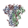
- EMDB-61600: Cryo-EM structure of SA-BatCoV (Neoromicia/PML-PHE1/RSA/2011) S-trimer -
+ Open data

- Basic information
| Entry |  | ||||||||||||
|---|---|---|---|---|---|---|---|---|---|---|---|---|---|
| Title | Cryo-EM structure of SA-BatCoV (Neoromicia/PML-PHE1/RSA/2011) S-trimer | ||||||||||||
 Map data Map data | |||||||||||||
 Sample Sample |
| ||||||||||||
 Keywords Keywords | spike protein / VIRAL PROTEIN | ||||||||||||
| Function / homology |  Function and homology information Function and homology informationvirion component / host cell endoplasmic reticulum-Golgi intermediate compartment membrane / receptor-mediated virion attachment to host cell / endocytosis involved in viral entry into host cell / fusion of virus membrane with host plasma membrane / fusion of virus membrane with host endosome membrane / viral envelope / host cell plasma membrane / virion membrane / membrane Similarity search - Function | ||||||||||||
| Biological species |  Coronavirus Neoromicia/PML-PHE1/RSA/2011 / Enterobacteria phage T4 (virus) Coronavirus Neoromicia/PML-PHE1/RSA/2011 / Enterobacteria phage T4 (virus) | ||||||||||||
| Method | single particle reconstruction / cryo EM / Resolution: 3.1 Å | ||||||||||||
 Authors Authors | Yuan H / Xiong X | ||||||||||||
| Funding support | 3 items
| ||||||||||||
 Citation Citation | Journal: Sci Adv / Year: 2025 Title: Structures and receptor binding activities of merbecovirus spike proteins reveal key signatures for human DPP4 adaptation. Authors: Hang Yuan / Jingjing Wang / Yong Ma / Zimu Li / Xijie Gao / Gul Habib / Banghui Liu / Jing Chen / Jun He / Peng Zhou / Zheng-Li Shi / Xinwen Chen / Xiaoli Xiong /  Abstract: Merbecoviruses from bats, pangolins, and hedgehogs pose significant zoonotic threats, with a limited understanding of receptor binding by their spike (S) proteins. Here, we report cryo-EM structures ...Merbecoviruses from bats, pangolins, and hedgehogs pose significant zoonotic threats, with a limited understanding of receptor binding by their spike (S) proteins. Here, we report cryo-EM structures of GD-BatCoV (BtCoV-422) and SE-PangolinCoV (MjHKU4r-CoV-1) RBDs in complex with human DPP4 (hDPP4). These structures exhibit a substantial offset in their hDPP4 interaction interfaces, revealing a conserved hydrophobic cluster as a convergent signature of DPP4 binding within the MERS-HKU4 clade of merbecoviruses. Structure-guided mutagenesis demonstrates that favorable interactions are distributed across multiple receptor binding motif (RBM) regions, working synergistically to confer high-affinity hDPP4 binding. Swapping of the merbecovirus RBM regions indicate limited plasticity and interchangeability among these regions. In addition, we report cryo-EM structures of six merbecovirus S-trimers. Structure-based phylogenetics suggests that hDPP4-binding merbecoviruses undergo convergent evolution, while ACE2-binding merbecoviruses exhibit diversification in their binding mechanisms. These findings offer critical insights into merbecovirus receptor utilization, providing a structural understanding for future surveillance. | ||||||||||||
| History |
|
- Structure visualization
| Supplemental images |
|---|

- Downloads & links
-EMDB archive
| Map data | emd_61600.map.gz | 49.5 MB | EMDB map data format | |
|---|---|---|---|---|
| Header (meta data) | emd-61600-v30.xmlemd-61600.xml | 20.3 KB 20.3 KB | Display Display | EMDB header |
| Images |  emd_61600.png emd_61600.png | 18.7 KB | ||
| Filedesc metadata | emd-61600.cif.gz | 7 KB | ||
| Others | emd_61600_half_map_1.map.gzemd_61600_half_map_2.map.gz | 40.8 MB 40.8 MB | ||
| Archive directory |  http://ftp.pdbj.org/pub/emdb/structures/EMD-61600ftp://ftp.pdbj.org/pub/emdb/structures/EMD-61600 http://ftp.pdbj.org/pub/emdb/structures/EMD-61600ftp://ftp.pdbj.org/pub/emdb/structures/EMD-61600 | HTTPS FTP |
-Related structure data
| Related structure data |  9jmfMC  9jmgC  9jmhC  9jmiC  9jmjC  9jmmC  9jmnC  9jmoC  9jmpC M: atomic model generated by this map C: citing same article ( |
|---|---|
| Similar structure data |


-Links
| EMDB pages | EMDB (EBI/PDBe) / EMDataResource |
|---|---|
| Related items in Molecule of the Month |


-Map
| File | Download / File: emd_61600.map.gz / Format: CCP4 / Size: 52.7 MB / Type: IMAGE STORED AS FLOATING POINT NUMBER (4 BYTES) | ||||||||||||||||||||||||||||||||||||
|---|---|---|---|---|---|---|---|---|---|---|---|---|---|---|---|---|---|---|---|---|---|---|---|---|---|---|---|---|---|---|---|---|---|---|---|---|---|
| Projections & slices | Image control
Images are generated by Spider. | ||||||||||||||||||||||||||||||||||||
| Voxel size | X=Y=Z: 1.32 Å | ||||||||||||||||||||||||||||||||||||
| Density |
| ||||||||||||||||||||||||||||||||||||
| Symmetry | Space group: 1 | ||||||||||||||||||||||||||||||||||||
| Details | EMDB XML:
|
 Z (Sec.)
 Y (Row.)
 X (Col.)


-Supplemental data
-Half map: #1
| File | emd_61600_half_map_1.map | ||||||||||||
|---|---|---|---|---|---|---|---|---|---|---|---|---|---|
| Projections & Slices |
| ||||||||||||
| Density Histograms |


-Half map: #2
| File | emd_61600_half_map_2.map | ||||||||||||
|---|---|---|---|---|---|---|---|---|---|---|---|---|---|
| Projections & Slices |
| ||||||||||||
| Density Histograms |


- Sample components
-Entire : SA-BatCoV (Neoromicia/PML-PHE1/RSA/2011) Spike protein
| Entire | Name: SA-BatCoV (Neoromicia/PML-PHE1/RSA/2011) Spike protein |
|---|---|
| Components |
|
-Supramolecule #1: SA-BatCoV (Neoromicia/PML-PHE1/RSA/2011) Spike protein
| Supramolecule | Name: SA-BatCoV (Neoromicia/PML-PHE1/RSA/2011) Spike protein type: organelle_or_cellular_component / ID: 1 / Parent: 0 / Macromolecule list: #1 |
|---|---|
| Source (natural) | Organism: Coronavirus Neoromicia/PML-PHE1/RSA/2011 |
| Molecular weight | Theoretical: 450 KDa |
-Macromolecule #1: Spike glycoprotein,Fibritin
| Macromolecule | Name: Spike glycoprotein,Fibritin / type: protein_or_peptide / ID: 1 / Number of copies: 3 / Enantiomer: LEVO |
|---|---|
| Source (natural) | Organism: Enterobacteria phage T4 (virus) |
| Molecular weight | Theoretical: 150.804984 KDa |
| Recombinant expression | Organism:  Homo sapiens (human) Homo sapiens (human) |
| Sequence | String: MTYSVFPLMC LLTFIGANAK IVTLPGNDAT GYCPSVDMQP SYFIQHNWPE PIDMNKADGV IYPNGRTYSN ITLQTTNLFP KNGDLGTQY VYSASNHKST ANDAFISNYS YYGNPFGDGI VIRIGQNANK TGSVIVGQAQ TTMKKIYPAL MLGSSFGNFS A NNKSGAYF ...String: MTYSVFPLMC LLTFIGANAK IVTLPGNDAT GYCPSVDMQP SYFIQHNWPE PIDMNKADGV IYPNGRTYSN ITLQTTNLFP KNGDLGTQY VYSASNHKST ANDAFISNYS YYGNPFGDGI VIRIGQNANK TGSVIVGQAQ TTMKKIYPAL MLGSSFGNFS A NNKSGAYF NHTLLILPSK CGTVFQVAYC LLQPRTESKC PGNSNYVSYF LADSPSDCSS TSDEIRRNGL RDIRKFFNLV NC TYFEEFN VTADERAEWF GIVQDAQGVH LYTSRKNGFN SNNLFLFATV PIYDKLNYYT VIPRSVITPS NQRDAWAAFY IYP LHQLSY LLNFDVNGYI TQAADCGYND YTQLICSYGD FNMKSGVYST SYYSAKPVGA YYEAHVYPDC NFTELFRERA PTIM QYKRE VFTRCNYNLS LLLSLVQVDE FVCDKITPEA LATGCYSSLT VDWFAFPYAW KSYLAIGSAD RIVRFNYNQD YSNPS CRIH SKVNSSIGIS YAGAYSYITN CNYGATNKDD VVKPGGRASQ QCITGALNSP TTGQLWAYNF GGVPYRVSRL TYTDHL SDP LDMVYVITVK YEPGAETVCP KQIRPDYSTN ITHLLGSCIS YDIYGITGTG VFQLCNATGI RQQKFVYDKF DNIIGFH SD DGNYYCVAPC VSVPVSVIYD DKTNQYATLF GSVACQHIST MAAQFSRETR ASLVSRNMQN LLQTSVGCVM GFHETNDT V EECHLSLGQS LCAIPPNTNL RSGRSTFGLG SLAYNSPLRV DALNSSEFKV SLPLNFTFGV TQEYIETSIQ KITVDCKQY VCNGFAKCEK LLEQYGQFCS KINQALHGAN LRQDDSVRNL FESVKTPQTV PLTTGFGGEF NLTLLEPLSV STGSSNARSA LEELLFDSV TIADPGYMQG YDDCMQQGPA SARDLICAQY VAGYKVLPPL MDVNMEAAYT SSLLGSIAGA GWTAGLSSFA A IPFAQSIF YRLNGVGITQ QVLSENQKII ANKFNQALGA MQTGFTTTNE AFQKVQDAVN TNAQALAKLA SELSNTFGAI SS SIGDIIQ RLDVLEQEVQ IDRLINGRLT TLNAFVAQQL VRSESAARSA QLAKDKVNEC VKSQSTRSGF CGQGTHIVSF VIN APNGLY FMHVGYHPSQ HIEVVAAYGL CDSANPTNCI APVNGYFIKN QTTRSADEWS YTGSSFYAPE PITTLNTRYV APQV TFQNI SNNLPPPLLS NSTGTDFKDE LDEFFKNVST NIPNFGALTQ INTTLLDLSG EMLALQEVVK ALNESYIDLK ELGNY TYYN KWPGSGYIPE APRDGQAYVR KDGEWVLLST FLLEVLFQGP GHHHHHHHHS AWSHPQFEKG GGSGGGGSGG SAWSHP QFE KSA UniProtKB: Spike glycoprotein, Fibritin |
-Macromolecule #3: 2-acetamido-2-deoxy-beta-D-glucopyranose
| Macromolecule | Name: 2-acetamido-2-deoxy-beta-D-glucopyranose / type: ligand / ID: 3 / Number of copies: 33 / Formula: NAG |
|---|---|
| Molecular weight | Theoretical: 221.208 Da |
| Chemical component information |  ChemComp-NAG: |
-Macromolecule #4: LINOLEIC ACID
| Macromolecule | Name: LINOLEIC ACID / type: ligand / ID: 4 / Number of copies: 3 / Formula: EIC |
|---|---|
| Molecular weight | Theoretical: 280.445 Da |
| Chemical component information |  ChemComp-EIC: |
-Experimental details
-Structure determination
| Method | cryo EM |
|---|---|
 Processing Processing | single particle reconstruction |
| Aggregation state | particle |
-Sample preparation
| Buffer | pH: 7.4 |
|---|---|
| Vitrification | Cryogen name: ETHANE |
- Electron microscopy
| Microscope | FEI TALOS ARCTICA |
|---|---|
| Image recording | Film or detector model: GATAN K3 (6k x 4k) / Average electron dose: 60.0 e/Å2 |
| Electron beam | Acceleration voltage: 200 kV / Electron source:  FIELD EMISSION GUN FIELD EMISSION GUN |
| Electron optics | Illumination mode: FLOOD BEAM / Imaging mode: OTHER / Nominal defocus max: 2.5 µm / Nominal defocus min: 0.8 µm |
| Experimental equipment |  Model: Talos Arctica / Image courtesy: FEI Company |