Movie
Movie Controller


+ Open data

- Basic information
| Entry |  | ||||||||||||
|---|---|---|---|---|---|---|---|---|---|---|---|---|---|
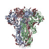
| Title | Cryo-EM structure of Japan-BatCoV (Vs-CoV-1) S-trimer | ||||||||||||
 Map data Map data | |||||||||||||
 Sample Sample |
| ||||||||||||
 Keywords Keywords | spike protein / VIRAL PROTEIN | ||||||||||||
| Function / homology |  Function and homology information Function and homology informationvirion component / host cell endoplasmic reticulum-Golgi intermediate compartment membrane / receptor-mediated virion attachment to host cell / endocytosis involved in viral entry into host cell / fusion of virus membrane with host plasma membrane / fusion of virus membrane with host endosome membrane / viral envelope / host cell plasma membrane / virion membrane / membrane Similarity search - Function | ||||||||||||
| Biological species |  Bat coronavirus / Tequatrovirus T4 Bat coronavirus / Tequatrovirus T4 | ||||||||||||
| Method | single particle reconstruction / cryo EM / Resolution: 2.8 Å | ||||||||||||
 Authors Authors | Yuan H / Xiong X | ||||||||||||
| Funding support | 3 items
| ||||||||||||
 Citation Citation | Journal: Sci Adv / Year: 2025 Title: Structures and receptor binding activities of merbecovirus spike proteins reveal key signatures for human DPP4 adaptation. Authors: Hang Yuan / Jingjing Wang / Yong Ma / Zimu Li / Xijie Gao / Gul Habib / Banghui Liu / Jing Chen / Jun He / Peng Zhou / Zheng-Li Shi / Xinwen Chen / Xiaoli Xiong /  Abstract: Merbecoviruses from bats, pangolins, and hedgehogs pose significant zoonotic threats, with a limited understanding of receptor binding by their spike (S) proteins. Here, we report cryo-EM structures ...Merbecoviruses from bats, pangolins, and hedgehogs pose significant zoonotic threats, with a limited understanding of receptor binding by their spike (S) proteins. Here, we report cryo-EM structures of GD-BatCoV (BtCoV-422) and SE-PangolinCoV (MjHKU4r-CoV-1) RBDs in complex with human DPP4 (hDPP4). These structures exhibit a substantial offset in their hDPP4 interaction interfaces, revealing a conserved hydrophobic cluster as a convergent signature of DPP4 binding within the MERS-HKU4 clade of merbecoviruses. Structure-guided mutagenesis demonstrates that favorable interactions are distributed across multiple receptor binding motif (RBM) regions, working synergistically to confer high-affinity hDPP4 binding. Swapping of the merbecovirus RBM regions indicate limited plasticity and interchangeability among these regions. In addition, we report cryo-EM structures of six merbecovirus S-trimers. Structure-based phylogenetics suggests that hDPP4-binding merbecoviruses undergo convergent evolution, while ACE2-binding merbecoviruses exhibit diversification in their binding mechanisms. These findings offer critical insights into merbecovirus receptor utilization, providing a structural understanding for future surveillance. | ||||||||||||
| History |
|
- Structure visualization
| Supplemental images |
|---|

- Downloads & links
-EMDB archive
| Map data | emd_61608.map.gz | 166.5 MB | EMDB map data format | |
|---|---|---|---|---|
| Header (meta data) | emd-61608-v30.xmlemd-61608.xml | 20 KB 20 KB | Display Display | EMDB header |
| Images |  emd_61608.png emd_61608.png | 14.9 KB | ||
| Filedesc metadata | emd-61608.cif.gz | 7.2 KB | ||
| Others | emd_61608_half_map_1.map.gzemd_61608_half_map_2.map.gz | 140.6 MB 140.6 MB | ||
| Archive directory |  http://ftp.pdbj.org/pub/emdb/structures/EMD-61608ftp://ftp.pdbj.org/pub/emdb/structures/EMD-61608 http://ftp.pdbj.org/pub/emdb/structures/EMD-61608ftp://ftp.pdbj.org/pub/emdb/structures/EMD-61608 | HTTPS FTP |
-Related structure data
| Related structure data |  9jmoMC  9jmfC  9jmgC  9jmhC  9jmiC  9jmjC  9jmmC  9jmnC  9jmpC M: atomic model generated by this map C: citing same article ( |
|---|---|
| Similar structure data |


-Links
| EMDB pages | EMDB (EBI/PDBe) / EMDataResource |
|---|---|
| Related items in Molecule of the Month |


-Map
| File | Download / File: emd_61608.map.gz / Format: CCP4 / Size: 178 MB / Type: IMAGE STORED AS FLOATING POINT NUMBER (4 BYTES) | ||||||||||||||||||||||||||||||||||||
|---|---|---|---|---|---|---|---|---|---|---|---|---|---|---|---|---|---|---|---|---|---|---|---|---|---|---|---|---|---|---|---|---|---|---|---|---|---|
| Projections & slices | Image control
Images are generated by Spider. | ||||||||||||||||||||||||||||||||||||
| Voxel size | X=Y=Z: 0.88 Å | ||||||||||||||||||||||||||||||||||||
| Density |
| ||||||||||||||||||||||||||||||||||||
| Symmetry | Space group: 1 | ||||||||||||||||||||||||||||||||||||
| Details | EMDB XML:
|
 Z (Sec.)
 Y (Row.)
 X (Col.)


-Supplemental data
-Half map: #2
| File | emd_61608_half_map_1.map | ||||||||||||
|---|---|---|---|---|---|---|---|---|---|---|---|---|---|
| Projections & Slices |
| ||||||||||||
| Density Histograms |


-Half map: #1
| File | emd_61608_half_map_2.map | ||||||||||||
|---|---|---|---|---|---|---|---|---|---|---|---|---|---|
| Projections & Slices |
| ||||||||||||
| Density Histograms |


- Sample components
-Entire : Japan-BatCoV (Vs-CoV-1) Spike protein
| Entire | Name: Japan-BatCoV (Vs-CoV-1) Spike protein |
|---|---|
| Components |
|
-Supramolecule #1: Japan-BatCoV (Vs-CoV-1) Spike protein
| Supramolecule | Name: Japan-BatCoV (Vs-CoV-1) Spike protein / type: organelle_or_cellular_component / ID: 1 / Parent: 0 / Macromolecule list: #1 |
|---|---|
| Source (natural) | Organism: Bat coronavirus |
-Macromolecule #1: Spike glycoprotein,Fibritin
| Macromolecule | Name: Spike glycoprotein,Fibritin / type: protein_or_peptide / ID: 1 / Number of copies: 3 / Enantiomer: LEVO |
|---|---|
| Source (natural) | Organism: Tequatrovirus T4 |
| Molecular weight | Theoretical: 151.802391 KDa |
| Recombinant expression | Organism:  Homo sapiens (human) Homo sapiens (human) |
| Sequence | String: MTHLMFLLMF LLTPIRGDVD LGPDSTRDTC DEVVVQVSAF QKDWPQPINP LFADGIIYPT GRSYSNISLS YQGLFPRQGD LGRQYIYSV THAGDSTLYP PQGIGKHFIS NYSAETLPFG NGFVVRIGFS ANKTGSLVIS PTTSKVIKKI YPAFMLGTAV T SFAHGHAG ...String: MTHLMFLLMF LLTPIRGDVD LGPDSTRDTC DEVVVQVSAF QKDWPQPINP LFADGIIYPT GRSYSNISLS YQGLFPRQGD LGRQYIYSV THAGDSTLYP PQGIGKHFIS NYSAETLPFG NGFVVRIGFS ANKTGSLVIS PTTSKVIKKI YPAFMLGTAV T SFAHGHAG RYLNHTLVIL PESCGTSLTV FYCILEPRNG TRCPNHQDYT SYVIWETPHL DCGSSVNRNA TLNSFKVSFN LR QCKFMRT YNISDDENQE WFGITQNAQG VHLYSSRKGD LYGGNMFLFD TLPVYETLKY YTVIPRSLRS RLNERQAWAA FYV YSLHKL TYLLDFSVDG YVRRAIDCGY DDVAQLFCSY ESFDVDSGVY SVSSFEAQPR GYFVEQPHSS ECDFSAMFSS QPPQ VYNFS RLVFTNCNYN LTRLLSLFQV SEFSCHQVSP SALASGCYSS LTVDYFAYPL SMASYLRQGS TGPIAQFNYK QDFTN PTCR ILAAVPSNVT IPLPQSYMWL TQCYTNSPSN SHYVEAGSYT PCLGLASRGF NTFNSMHYDP ATGLTATGSY QYISEE ALE MAFIISVQYG TDSNSVCPLQ AVHNDTKVDD KLGRCIEYSL YGVTGRGVFQ NCTPVGLKQQ RFVYDGFDNL VGYHSDN GN YYCVRPCVSV PVSVIYDKAT NNHATLFGSV ACEHVDTMMS QFSRMTQSRL RVRNIDDAGP MQTSVGCVVG LVNTSLIV D NCQLPLGQSL CAVPATTIRA ATGTTSSLQL ATINFTEPHM LTPINSSRFI VEIPTNFSFG VTQEFIQTTI QKVTVDCKQ YVCNGFEKCE QLLREYGQFC AKINQALHGA NLRQDESVSN LFSNIKTQAT QPLDAGLTGD FNLTLLQVPK VVTSQYSYRS AIEDLLFNK VTIADPGYMQ GYDECMKQGP PSARDLICAQ FVAGYKVLPP LYDPNMESMY TTSLTGAILG AAWTAGASSF A AIPFAQSV FYRMNGIGIT QQVLSENQKQ IANKFNQALG AMQTGFTTTN LAFAKVQEAV NANAQALSKL ASELSNTFGA IS SSISDIL KRLDAVEQEA QIDRLINGRL TSLNAFVAQQ LVRSETAARS SQLAVDKVNE CVKSQSKRNG FCGSGNHIVS FVS NAPNGL YFFHVGYVPT AHINATAAYG LCNTELPPRC IAPIDGYFVT NSSATRDADP EWYYTGSAFY NPEPITLANT RYVS KDTKY QELNNKLPPP LLSNQTDESF RDELEEFFKN VSSQGPNFQE ISKINATLLD LSDEMKVLNE VVKQLNESYI DLKEL GNYT YYQKWPGSGY IPEAPRDGQA YVRKDGEWVL LSTFLLEVLF QGPGHHHHHH HHSAWSHPQF EKGGGSGGGG SGGSAW SHP QFEKSA UniProtKB: Spike glycoprotein, Fibritin |
-Macromolecule #4: 2-acetamido-2-deoxy-beta-D-glucopyranose
| Macromolecule | Name: 2-acetamido-2-deoxy-beta-D-glucopyranose / type: ligand / ID: 4 / Number of copies: 31 / Formula: NAG |
|---|---|
| Molecular weight | Theoretical: 221.208 Da |
| Chemical component information |  ChemComp-NAG: |
-Macromolecule #5: LINOLEIC ACID
| Macromolecule | Name: LINOLEIC ACID / type: ligand / ID: 5 / Number of copies: 3 / Formula: EIC |
|---|---|
| Molecular weight | Theoretical: 280.445 Da |
| Chemical component information |  ChemComp-EIC: |
-Experimental details
-Structure determination
| Method | cryo EM |
|---|---|
 Processing Processing | single particle reconstruction |
| Aggregation state | particle |
-Sample preparation
| Buffer | pH: 7.4 |
|---|---|
| Vitrification | Cryogen name: ETHANE |
- Electron microscopy
| Microscope | FEI TALOS ARCTICA |
|---|---|
| Image recording | Film or detector model: GATAN K3 (6k x 4k) / Average electron dose: 60.0 e/Å2 |
| Electron beam | Acceleration voltage: 200 kV / Electron source:  FIELD EMISSION GUN FIELD EMISSION GUN |
| Electron optics | Illumination mode: OTHER / Imaging mode: BRIGHT FIELD / Nominal defocus max: 2.5 µm / Nominal defocus min: 0.8 µm |
| Experimental equipment |  Model: Talos Arctica / Image courtesy: FEI Company |