 Movie
Movie Controller

[English] 日本語
 Yorodumi
- EMDB-5967: Single particle EM reveals plasticity of interactions between the... -
+ Open data

- Basic information
| Entry | Database: EMDB / ID: EMD-5967 | |||||||||
|---|---|---|---|---|---|---|---|---|---|---|
| Title | Single particle EM reveals plasticity of interactions between the adenovirus penton base and integrin alphaV-beta3 | |||||||||
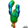
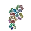
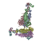
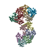
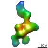
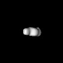
 Map data Map data | Reconstruction of the human alphaV-beta3 integrin in complex with the monomeric RGD-loop containing insertion domain of the adenovirus 9 penton base subunit | |||||||||
 Sample Sample |
| |||||||||
 Keywords Keywords | adenovirus / integrin / virus infection / electron microscopy / fluorescence correlation microscopy | |||||||||
| Function / homology |  Function and homology information Function and homology informationT=25 icosahedral viral capsid / integrin alphav-beta8 complex / integrin alphav-beta6 complex / transforming growth factor beta production / negative regulation of entry of bacterium into host cell / integrin alphav-beta5 complex / opsonin binding / integrin alphav-beta1 complex / Cross-presentation of particulate exogenous antigens (phagosomes) / extracellular matrix protein binding ...T=25 icosahedral viral capsid / integrin alphav-beta8 complex / integrin alphav-beta6 complex / transforming growth factor beta production / negative regulation of entry of bacterium into host cell / integrin alphav-beta5 complex / opsonin binding / integrin alphav-beta1 complex / Cross-presentation of particulate exogenous antigens (phagosomes) / extracellular matrix protein binding / Laminin interactions / integrin alphav-beta3 complex / negative regulation of lipoprotein metabolic process / alphav-beta3 integrin-PKCalpha complex / positive regulation of small GTPase mediated signal transduction / entry into host cell by a symbiont-containing vacuole / alphav-beta3 integrin-HMGB1 complex / negative regulation of lipid transport / regulation of phagocytosis / Elastic fibre formation / alphav-beta3 integrin-IGF-1-IGF1R complex / transforming growth factor beta binding / filopodium membrane / extracellular matrix binding / apolipoprotein A-I-mediated signaling pathway / negative regulation of low-density lipoprotein particle clearance / wound healing, spreading of epidermal cells / apoptotic cell clearance / integrin complex / cell adhesion mediated by integrin / Molecules associated with elastic fibres / heterotypic cell-cell adhesion / negative chemotaxis / Mechanical load activates signaling by PIEZO1 and integrins in osteocytes / Syndecan interactions / positive regulation of osteoblast proliferation / microvillus membrane / cell-substrate adhesion / PECAM1 interactions / endodermal cell differentiation / TGF-beta receptor signaling activates SMADs / lamellipodium membrane / fibronectin binding / positive regulation of intracellular signal transduction / negative regulation of macrophage derived foam cell differentiation / negative regulation of lipid storage / ECM proteoglycans / Integrin cell surface interactions / vasculogenesis / specific granule membrane / coreceptor activity / ERK1 and ERK2 cascade / phagocytic vesicle / extrinsic apoptotic signaling pathway in absence of ligand / substrate adhesion-dependent cell spreading / positive regulation of cell adhesion / Turbulent (oscillatory, disturbed) flow shear stress activates signaling by PIEZO1 and integrins in endothelial cells / cell-matrix adhesion / protein kinase C binding / negative regulation of extrinsic apoptotic signaling pathway / Signal transduction by L1 / integrin-mediated signaling pathway / cell-cell adhesion / VEGFA-VEGFR2 Pathway / integrin binding / ruffle membrane / calcium ion transmembrane transport / cell migration / virus receptor activity / positive regulation of cytosolic calcium ion concentration / signaling receptor activity / protease binding / angiogenesis / cell adhesion / positive regulation of cell migration / endocytosis involved in viral entry into host cell / external side of plasma membrane / focal adhesion / positive regulation of cell population proliferation / Neutrophil degranulation / symbiont entry into host cell / virion attachment to host cell / cell surface / extracellular exosome / membrane / metal ion binding / plasma membrane / cytosol Similarity search - Function | |||||||||
| Biological species |  Homo sapiens (human) / Homo sapiens (human) /  Human adenovirus 9 Human adenovirus 9 | |||||||||
| Method | single particle reconstruction / negative staining / Resolution: 53.0 Å | |||||||||
 Authors Authors | Veesler D / Cupelli K / Burger M / Graber P / Stehle T / Johnson JE | |||||||||
 Citation Citation | Journal: Proc Natl Acad Sci U S A / Year: 2014 Title: Single-particle EM reveals plasticity of interactions between the adenovirus penton base and integrin αVβ3. Authors: David Veesler / Karolina Cupelli / Markus Burger / Peter Gräber / Thilo Stehle / John E Johnson /   Abstract: Human adenoviruses are double-stranded DNA viruses responsible for numerous infections, some of which can be fatal. Furthermore, adenoviruses are currently used in clinical trials as vectors for gene ...Human adenoviruses are double-stranded DNA viruses responsible for numerous infections, some of which can be fatal. Furthermore, adenoviruses are currently used in clinical trials as vectors for gene therapy applications. Although initial binding of adenoviruses to host attachment receptors has been extensively characterized, the interactions with the entry receptor (integrins) remain poorly understood at the structural level. We characterized the interactions between the adenovirus 9 penton base subunit and αVβ3 integrin using fluorescence correlation spectroscopy and single-particle electron microscopy to understand the mechanisms underlying virus internalization and infection. Our results indicate that the penton base subunit can bind integrins with high affinity and in several different orientations. These outcomes correlate with the requirement of the pentameric penton base to simultaneously bind several integrins to enable their clustering and promote virus entry into the host cell. | |||||||||
| History |
|
- Structure visualization
| Movie |
Movie viewer |
|---|---|
| Structure viewer | EM map: SurfViewMolmilJmol/JSmol |
| Supplemental images |
 UCSF Chimera
- Downloads & links
-EMDB archive
| Map data | emd_5967.map.gz | 2.1 MB | EMDB map data format | |
|---|---|---|---|---|
| Header (meta data) | emd-5967-v30.xmlemd-5967.xml | 10.8 KB 10.8 KB | Display Display | EMDB header |
| Images |  400_5967.gif 400_5967.gif 80_5967.gif 80_5967.gif | 18.4 KB 2.3 KB | ||
| Archive directory |  http://ftp.pdbj.org/pub/emdb/structures/EMD-5967ftp://ftp.pdbj.org/pub/emdb/structures/EMD-5967 http://ftp.pdbj.org/pub/emdb/structures/EMD-5967ftp://ftp.pdbj.org/pub/emdb/structures/EMD-5967 | HTTPS FTP |
-Related structure data
| Related structure data |  5955C 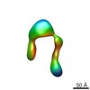 5956C 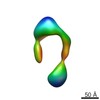 5957C  5958C 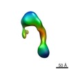 5959C 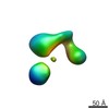 5960C 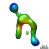 5961C 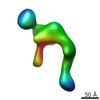 5962C 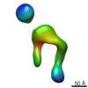 5963C 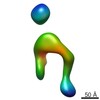 5964C 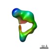 5965C 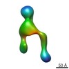 5966C 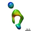 5968C 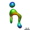 5969C 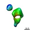 5970C 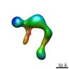 5971C 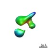 5972C 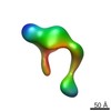 5973C C: citing same article ( |
|---|---|
| Similar structure data |
-Links
| EMDB pages | EMDB (EBI/PDBe) / EMDataResource |
|---|---|
| Related items in Molecule of the Month |


-Map
| File | Download / File: emd_5967.map.gz / Format: CCP4 / Size: 7.8 MB / Type: IMAGE STORED AS FLOATING POINT NUMBER (4 BYTES) | ||||||||||||||||||||||||||||||||||||||||||||||||||||||||||||||||||||
|---|---|---|---|---|---|---|---|---|---|---|---|---|---|---|---|---|---|---|---|---|---|---|---|---|---|---|---|---|---|---|---|---|---|---|---|---|---|---|---|---|---|---|---|---|---|---|---|---|---|---|---|---|---|---|---|---|---|---|---|---|---|---|---|---|---|---|---|---|---|
| Annotation | Reconstruction of the human alphaV-beta3 integrin in complex with the monomeric RGD-loop containing insertion domain of the adenovirus 9 penton base subunit | ||||||||||||||||||||||||||||||||||||||||||||||||||||||||||||||||||||
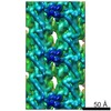
| Projections & slices | Image control
Images are generated by Spider. | ||||||||||||||||||||||||||||||||||||||||||||||||||||||||||||||||||||
| Voxel size | X=Y=Z: 4.1 Å | ||||||||||||||||||||||||||||||||||||||||||||||||||||||||||||||||||||
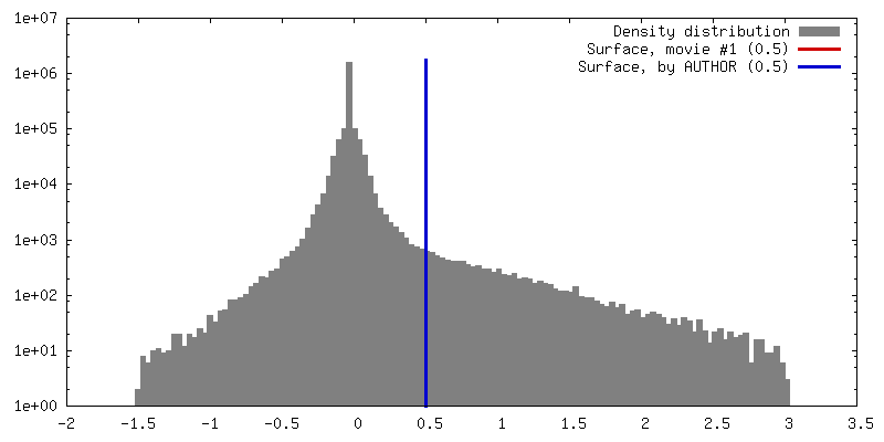
| Density |
| ||||||||||||||||||||||||||||||||||||||||||||||||||||||||||||||||||||
| Symmetry | Space group: 1 | ||||||||||||||||||||||||||||||||||||||||||||||||||||||||||||||||||||
| Details | EMDB XML:
CCP4 map header:
| ||||||||||||||||||||||||||||||||||||||||||||||||||||||||||||||||||||
 Z (Sec.)
 Y (Row.)
 X (Col.)


-Supplemental data
- Sample components
-Entire : Human alphaV-beta3 integrin ectodomain in complex with the monome...
| Entire | Name: Human alphaV-beta3 integrin ectodomain in complex with the monomeric RGD-loop containing insertion domain of the adenovirus 9 penton base subunit |
|---|---|
| Components |
|
-Supramolecule #1000: Human alphaV-beta3 integrin ectodomain in complex with the monome...
| Supramolecule | Name: Human alphaV-beta3 integrin ectodomain in complex with the monomeric RGD-loop containing insertion domain of the adenovirus 9 penton base subunit type: sample / ID: 1000 / Oligomeric state: heterotrimer / Number unique components: 2 |
|---|---|
| Molecular weight | Theoretical: 210 KDa |
-Macromolecule #1: alphaV-beta3 integrin ectodomain
| Macromolecule | Name: alphaV-beta3 integrin ectodomain / type: protein_or_peptide / ID: 1 / Details: Second UniProt identifier: P05106 / Number of copies: 1 / Oligomeric state: heterodimer / Recombinant expression: Yes |
|---|---|
| Source (natural) | Organism: Homo sapiens (human) / synonym: Human |
| Molecular weight | Theoretical: 180 KDa |
| Recombinant expression | Organism:   Spodoptera frugiperda (fall armyworm) / Recombinant cell: Sf9 / Recombinant plasmid: pFastBacDual Spodoptera frugiperda (fall armyworm) / Recombinant cell: Sf9 / Recombinant plasmid: pFastBacDual |
| Sequence | UniProtKB: Integrin alpha-V |
-Macromolecule #2: adenovirus 9 penton base
| Macromolecule | Name: adenovirus 9 penton base / type: protein_or_peptide / ID: 2 Details: Only the monomeric RGD-loop containing insertion domain of the adenovirus 9 penton base subunit is present in the construct used (residues 116 to 360). Number of copies: 1 / Oligomeric state: Monomer / Recombinant expression: Yes |
|---|---|
| Source (natural) | Organism: Human adenovirus 9 |
| Molecular weight | Theoretical: 30 KDa |
| Recombinant expression | Organism:  |
| Sequence | UniProtKB: Penton base |
-Experimental details
-Structure determination
| Method | negative staining |
|---|---|
 Processing Processing | single particle reconstruction |
| Aggregation state | particle |
-Sample preparation
| Buffer | pH: 7.5 / Details: 10 mM Tris, 150 mM NaCl, 2 mM MnCl2 |
|---|---|
| Staining | Type: NEGATIVE / Details: 2% uranyl formate |
| Grid | Details: C-flat 2/0.5 grids overlaid with a thin layer of carbon |
| Vitrification | Cryogen name: NONE / Instrument: OTHER |
- Electron microscopy
| Microscope | FEI TECNAI 12 |
|---|---|
| Date | May 30, 2013 |
| Image recording | Category: CCD / Film or detector model: TVIPS TEMCAM-F416 (4k x 4k) / Number real images: 1600 / Average electron dose: 24 e/Å2 |
| Electron beam | Acceleration voltage: 120 kV / Electron source: LAB6 |
| Electron optics | Illumination mode: FLOOD BEAM / Imaging mode: BRIGHT FIELD / Cs: 2.2 mm / Nominal defocus max: 2.8 µm / Nominal defocus min: 0.1 µm / Nominal magnification: 76097 |
| Sample stage | Specimen holder model: OTHER |
-Image processing
| Details | Random conical tilt reconstruction |
|---|---|
| Final reconstruction | Algorithm: OTHER / Resolution.type: BY AUTHOR / Resolution: 53.0 Å / Resolution method: OTHER / Software - Name: Spider / Number images used: 515 |
| Final two d classification | Number classes: 1 |
