Movie
Movie Controller

[English] 日本語
 Yorodumi
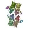
- EMDB-2005: Subnanometer reconstruction of GTPgammaS microtubules decorated w... -
+ Open data

- Basic information
| Entry | Database: EMDB / ID: EMD-2005 | |||||||||
|---|---|---|---|---|---|---|---|---|---|---|
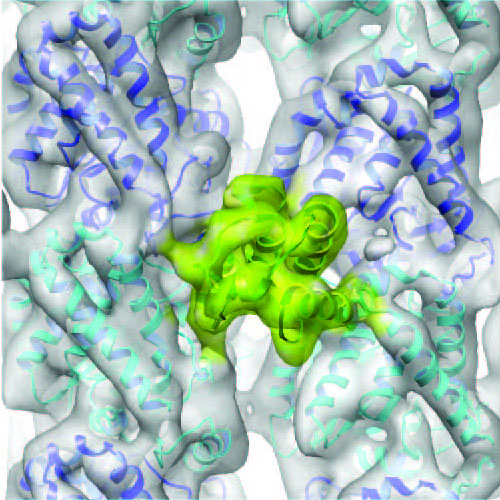
| Title | Subnanometer reconstruction of GTPgammaS microtubules decorated with monomeric Mal3 | |||||||||
 Map data Map data | Subnanometer reconstruction of GTPgammaS microtubules decorated with monomeric Mal3 | |||||||||
 Sample Sample |
| |||||||||
 Keywords Keywords | cytoskeleton / GTPase / end binding / calponin homology | |||||||||
| Function / homology |  Function and homology information Function and homology informationdynein-driven meiotic oscillatory nuclear movement / post-anaphase array microtubule end / nuclear migration involved in conjugation with cellular fusion / cortical microtubule / cell cortex of cell tip / karyogamy involved in conjugation with cellular fusion / mitotic spindle astral microtubule / mitotic spindle midzone / mitotic spindle pole body / nuclear microtubule ...dynein-driven meiotic oscillatory nuclear movement / post-anaphase array microtubule end / nuclear migration involved in conjugation with cellular fusion / cortical microtubule / cell cortex of cell tip / karyogamy involved in conjugation with cellular fusion / mitotic spindle astral microtubule / mitotic spindle midzone / mitotic spindle pole body / nuclear microtubule / protein localization to microtubule / astral microtubule / microtubule plus-end / cytoskeletal adaptor activity / attachment of mitotic spindle microtubules to kinetochore / microtubule plus-end binding / microtubule lateral binding / spindle midzone / ATPase activator activity / microtubule organizing center / regulation of microtubule polymerization or depolymerization / cytoplasmic microtubule / spindle assembly / molecular condensate scaffold activity / structural constituent of cytoskeleton / microtubule cytoskeleton organization / neuron migration / mitotic cell cycle / microtubule cytoskeleton / microtubule binding / microtubule / Hydrolases; Acting on acid anhydrides; Acting on GTP to facilitate cellular and subcellular movement / cell division / hydrolase activity / GTPase activity / GTP binding / metal ion binding / nucleus / cytoplasm Similarity search - Function | |||||||||
| Biological species |   | |||||||||
| Method | single particle reconstruction / cryo EM / Resolution: 8.6 Å | |||||||||
 Authors Authors | Maurer SP / Fourniol FJ / Bohner G / Moores CA / Surrey T | |||||||||
 Citation Citation | Journal: Cell / Year: 2012 Title: EBs recognize a nucleotide-dependent structural cap at growing microtubule ends. Authors: Sebastian P Maurer / Franck J Fourniol / Gergő Bohner / Carolyn A Moores / Thomas Surrey /  Abstract: Growing microtubule ends serve as transient binding platforms for essential proteins that regulate microtubule dynamics and their interactions with cellular substructures. End-binding proteins (EBs) ...Growing microtubule ends serve as transient binding platforms for essential proteins that regulate microtubule dynamics and their interactions with cellular substructures. End-binding proteins (EBs) autonomously recognize an extended region at growing microtubule ends with unknown structural characteristics and then recruit other factors to the dynamic end structure. Using cryo-electron microscopy, subnanometer single-particle reconstruction, and fluorescence imaging, we present a pseudoatomic model of how the calponin homology (CH) domain of the fission yeast EB Mal3 binds to the end regions of growing microtubules. The Mal3 CH domain bridges protofilaments except at the microtubule seam. By binding close to the exchangeable GTP-binding site, the CH domain is ideally positioned to sense the microtubule's nucleotide state. The same microtubule-end region is also a stabilizing structural cap protecting the microtubule from depolymerization. This insight supports a common structural link between two important biological phenomena, microtubule dynamic instability and end tracking. | |||||||||
| History |
|
- Structure visualization
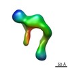
| Movie |
Movie viewer |
|---|---|
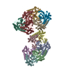
| Structure viewer | EM map: SurfViewMolmilJmol/JSmol |
| Supplemental images |
- Downloads & links
-EMDB archive
| Map data | emd_2005.map.gz | 815.4 KB | EMDB map data format | |
|---|---|---|---|---|
| Header (meta data) | emd-2005-v30.xmlemd-2005.xml | 12.9 KB 12.9 KB | Display Display | EMDB header |
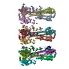
| Images |  2005-mal3-GTPgS.jpg 2005-mal3-GTPgS.jpg | 694.2 KB | ||
| Archive directory |  http://ftp.pdbj.org/pub/emdb/structures/EMD-2005ftp://ftp.pdbj.org/pub/emdb/structures/EMD-2005 http://ftp.pdbj.org/pub/emdb/structures/EMD-2005ftp://ftp.pdbj.org/pub/emdb/structures/EMD-2005 | HTTPS FTP |
-Related structure data
| Related structure data |  4aboMC 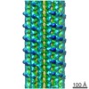 2004C 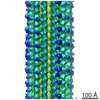 2006C M: atomic model generated by this map C: citing same article ( |
|---|---|
| Similar structure data |

-Links
| EMDB pages | EMDB (EBI/PDBe) / EMDataResource |
|---|---|
| Related items in Molecule of the Month |


-Map
| File | Download / File: emd_2005.map.gz / Format: CCP4 / Size: 859.4 KB / Type: IMAGE STORED AS FLOATING POINT NUMBER (4 BYTES) | ||||||||||||||||||||||||||||||||||||||||||||||||||||||||||||||||||||
|---|---|---|---|---|---|---|---|---|---|---|---|---|---|---|---|---|---|---|---|---|---|---|---|---|---|---|---|---|---|---|---|---|---|---|---|---|---|---|---|---|---|---|---|---|---|---|---|---|---|---|---|---|---|---|---|---|---|---|---|---|---|---|---|---|---|---|---|---|---|
| Annotation | Subnanometer reconstruction of GTPgammaS microtubules decorated with monomeric Mal3 | ||||||||||||||||||||||||||||||||||||||||||||||||||||||||||||||||||||
| Projections & slices | Image control
Images are generated by Spider. generated in cubic-lattice coordinate | ||||||||||||||||||||||||||||||||||||||||||||||||||||||||||||||||||||
| Voxel size | X=Y=Z: 2.2 Å | ||||||||||||||||||||||||||||||||||||||||||||||||||||||||||||||||||||
| Density |
| ||||||||||||||||||||||||||||||||||||||||||||||||||||||||||||||||||||
| Symmetry | Space group: 1 | ||||||||||||||||||||||||||||||||||||||||||||||||||||||||||||||||||||
| Details | EMDB XML:
CCP4 map header:
| ||||||||||||||||||||||||||||||||||||||||||||||||||||||||||||||||||||
 Z (Sec.)
 Y (Row.)
 X (Col.)


-Supplemental data
- Sample components
-Entire : GTPgammaS microtubules decorated with monomeric Mal3
| Entire | Name: GTPgammaS microtubules decorated with monomeric Mal3 |
|---|---|
| Components |
|
-Supramolecule #1000: GTPgammaS microtubules decorated with monomeric Mal3
| Supramolecule | Name: GTPgammaS microtubules decorated with monomeric Mal3 / type: sample / ID: 1000 Details: tubulin was mixed with GMPCPP microtubule seeds, GTPgammaS and monomeric Mal3, and incubated 3-10min at 37degC Oligomeric state: 13-protofilament microtubule / Number unique components: 4 |
|---|
-Macromolecule #1: Alpha tubulin
| Macromolecule | Name: Alpha tubulin / type: protein_or_peptide / ID: 1 / Name.synonym: Alpha tubulin dimer / Oligomeric state: Dimer / Recombinant expression: No / Database: NCBI |
|---|---|
| Source (natural) | Organism: |
| Molecular weight | Experimental: 50 KDa / Theoretical: 50 KDa |
-Macromolecule #2: beta tubulin
| Macromolecule | Name: beta tubulin / type: protein_or_peptide / ID: 2 / Name.synonym: beta tubulin / Oligomeric state: Dimer / Recombinant expression: No / Database: NCBI |
|---|---|
| Source (natural) | Organism: |
| Molecular weight | Experimental: 50 KDa / Theoretical: 50 KDa |
-Macromolecule #4: Mal3
| Macromolecule | Name: Mal3 / type: protein_or_peptide / ID: 4 / Name.synonym: Mal3 / Number of copies: 1 / Oligomeric state: monomer / Recombinant expression: Yes |
|---|---|
| Source (natural) | Organism: |
| Molecular weight | Experimental: 16 KDa / Theoretical: 16 KDa |
| Recombinant expression | Organism:  |
-Macromolecule #3: guanosine 5'-O-(gamma-thio)triphosphate
| Macromolecule | Name: guanosine 5'-O-(gamma-thio)triphosphate / type: ligand / ID: 3 / Name.synonym: GTPgammaS / Recombinant expression: No / Database: NCBI |
|---|---|
| Source (natural) | Organism: synthetic construct (others) |
-Experimental details
-Structure determination
| Method | cryo EM |
|---|---|
 Processing Processing | single particle reconstruction |
| Aggregation state | particle |
-Sample preparation
| Buffer | pH: 6.8 / Details: 40mM Pipes, 1mM MgCl2, 1mM EGTA |
|---|---|
| Grid | Details: 300 mesh lacey carbon grid |
| Vitrification | Cryogen name: ETHANE / Chamber humidity: 90 % / Instrument: OTHER / Details: Vitrification instrument: Vitrobot (FEI) / Method: Chamber at 37 degrees C, blot 2s |
- Electron microscopy
| Microscope | FEI TECNAI F20 |
|---|---|
| Temperature | Min: 88 K / Max: 98 K / Average: 93 K |
| Alignment procedure | Legacy - Astigmatism: Objective lens astigmatism was corrected at 150,000 times magnification |
| Image recording | Category: CCD / Film or detector model: GATAN ULTRASCAN 4000 (4k x 4k) / Number real images: 162 / Average electron dose: 17 e/Å2 / Details: sampling size 2.2 A per pixel |
| Electron beam | Acceleration voltage: 200 kV / Electron source:  FIELD EMISSION GUN FIELD EMISSION GUN |
| Electron optics | Illumination mode: FLOOD BEAM / Imaging mode: BRIGHT FIELD / Cs: 2.0 mm / Nominal defocus max: 3.6 µm / Nominal defocus min: 0.7 µm / Nominal magnification: 68000 |
| Sample stage | Specimen holder: Eucentric / Specimen holder model: OTHER |
| Experimental equipment |  Model: Tecnai F20 / Image courtesy: FEI Company |
-Image processing
| CTF correction | Details: FREALIGN |
|---|---|
| Final reconstruction | Algorithm: OTHER / Resolution.type: BY AUTHOR / Resolution: 8.6 Å / Resolution method: FSC 0.5 CUT-OFF / Software - Name: SPIDER, FREALIGN Details: approximately 129000 tubulin dimers were averaged together in the final map Number images used: 129000 |
-Atomic model buiding 1
| Initial model | PDB ID: |
|---|---|
| Software | Name: UCSF Chimera |
| Details | Protocol: Rigid body. Each tubulin monomer and a homology model for Mal3 CH domain were separately fitted using automatic fitting, and the multi-subunit fitting was refined in Flex-EM |
| Refinement | Space: REAL / Protocol: RIGID BODY FIT / Target criteria: cross-correlation |
| Output model | PDB-4abo: |