 Movie
Movie Controller

[English] 日本語
 Yorodumi
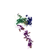
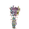
- PDB-6w4q: Crystal structure of full-length tailspike protein 2 (TSP2, ORF21... -
+ Open data

- Basic information
| Entry | Database: PDB / ID: 6w4q | ||||||
|---|---|---|---|---|---|---|---|
| Title | Crystal structure of full-length tailspike protein 2 (TSP2, ORF211) ) from Escherichia coli O157:H7 bacteriophage CAB120 | ||||||
 Components Components | Tail fiber | ||||||
 Keywords Keywords | HYDROLASE / glycosidase / tailspike protein / phage CBA120 / lipopolysaccharide degradation | ||||||
| Function / homology | : / Phage tail protein small four-stranded beta-sheet domain / metal ion binding / CARBONATE ION / Putative tail fiber Function and homology information Function and homology information | ||||||
| Biological species |  Escherichia virus CBA120 Escherichia virus CBA120 | ||||||
| Method |  X-RAY DIFFRACTION / SYNCHROTRON / MOLECULAR REPLACEMENT / molecular replacement / Resolution: 1.9 Å X-RAY DIFFRACTION / SYNCHROTRON / MOLECULAR REPLACEMENT / molecular replacement / Resolution: 1.9 Å | ||||||
 Authors Authors | Greenfield, J. / Herzberg, O. | ||||||
| Funding support |  United States, 1items United States, 1items
| ||||||
 Citation Citation | Journal: Sci Rep / Year: 2020 Title: Structure and function of bacteriophage CBA120 ORF211 (TSP2), the determinant of phage specificity towards E. coli O157:H7. Authors: Greenfield, J. / Shang, X. / Luo, H. / Zhou, Y. / Linden, S.B. / Heselpoth, R.D. / Leiman, P.G. / Nelson, D.C. / Herzberg, O. #1: Journal: PloS One / Year: 2014Title: Crystal structure of ORF210 from E. coli O157:H1 phage CBA120 (TSP1), a putative tailspike protein Authors: Chen, C. / Bales, P. / Greenfield, J. / Heselpoth, R.D. / Nelson, D.C. / Herzberg, O. #2: Journal: Sci Rep / Year: 2019Title: Structure and tailspike glycosidase machinery of ORF212 from E. coli O157:H7 phage CBA120 (TSP3). Authors: Greenfield, J. / Shang, X. / Luo, H. / Zhou, Y. / Heselpoth, R.D. / Nelson, D.C. / Herzberg, O. | ||||||
| History |
|
- Structure visualization
| Structure viewer | Molecule: MolmilJmol/JSmol |
|---|
- Downloads & links
-Download
| PDBx/mmCIF format | 6w4q.cif.gz | 966.5 KB | Display | PDBx/mmCIF format |
|---|---|---|---|---|
| PDB format | pdb6w4q.ent.gz | 775.3 KB | Display | PDB format |
| PDBx/mmJSON format | 6w4q.json.gz | Tree view | PDBx/mmJSON format | |
| Others |  Other downloads Other downloads |
-Validation report
| Arichive directory | https://data.pdbj.org/pub/pdb/validation_reports/w4/6w4qftp://data.pdbj.org/pub/pdb/validation_reports/w4/6w4q | HTTPS FTP |
|---|
-Related structure data
| Related structure data | 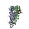 5w6pS S: Starting model for refinement |
|---|---|
| Similar structure data |


-Links
 PDBj
PDBj


- Assembly
| Deposited unit | 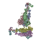
| ||||||||
|---|---|---|---|---|---|---|---|---|---|
| 1 | 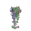
| ||||||||
| 2 | 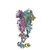
| ||||||||
| Unit cell |
|
-Components
-Protein , 1 types, 6 molecules ABCDEF
| #1: Protein | Mass: 99127.453 Da / Num. of mol.: 6 Source method: isolated from a genetically manipulated source Source: (gene. exp.) Escherichia virus CBA120 / Gene: orf211 / Plasmid: PBAD24Production host:  Strain (production host): rosetta-gami 2 / Variant (production host): DE3 / References: UniProt: G3M190 |
|---|
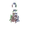
-Non-polymers , 6 types, 4004 molecules 


| #2: Chemical | ChemComp-GOL /  Mass: 92.094 Da / Num. of mol.: 78 / Source method: obtained synthetically / Formula: C3H8O3 Mass: 92.094 Da / Num. of mol.: 78 / Source method: obtained synthetically / Formula: C3H8O3#3: Chemical | ChemComp-SO4 /  Mass: 96.063 Da / Num. of mol.: 25 / Source method: obtained synthetically / Formula: SO4 Mass: 96.063 Da / Num. of mol.: 25 / Source method: obtained synthetically / Formula: SO4#4: Chemical | ChemComp-CL /  Mass: 35.453 Da / Num. of mol.: 7 / Source method: obtained synthetically / Formula: Cl Mass: 35.453 Da / Num. of mol.: 7 / Source method: obtained synthetically / Formula: Cl#5: Chemical |  Mass: 60.009 Da / Num. of mol.: 2 / Source method: obtained synthetically / Formula: CO3 Mass: 60.009 Da / Num. of mol.: 2 / Source method: obtained synthetically / Formula: CO3#6: Chemical | ChemComp-EDO /  Mass: 62.068 Da / Num. of mol.: 5 / Source method: obtained synthetically / Formula: C2H6O2 Mass: 62.068 Da / Num. of mol.: 5 / Source method: obtained synthetically / Formula: C2H6O2#7: Water | ChemComp-HOH / | Mass: 18.015 Da / Num. of mol.: 3887 / Source method: isolated from a natural source / Formula: H2O |
|---|
-Details
| Has ligand of interest | N |
|---|
-Experimental details
-Experiment
| Experiment | Method: X-RAY DIFFRACTION / Number of used crystals: 1 |
|---|
- Sample preparation
| Crystal | Density Matthews: 2.44 Å3/Da / Density % sol: 49.52 % |
|---|---|
| Crystal grow | Temperature: 298 K / Method: vapor diffusion, sitting drop / pH: 7 / Details: 0.8 M ammonium sulfate and 0.1M HEPES (pH 7.0) / Temp details: room temperature |
-Data collection
| Diffraction | Mean temperature: 100 K / Serial crystal experiment: N |
|---|---|
| Diffraction source | Source: SYNCHROTRON / Site: APS / Beamline: 23-ID-B / Wavelength: 1.0332 Å |
| Detector | Type: MARMOSAIC 300 mm CCD / Detector: CCD / Date: Nov 17, 2015 |
| Radiation | Monochromator: SI(111) / Protocol: SINGLE WAVELENGTH / Monochromatic (M) / Laue (L): M / Scattering type: x-ray |
| Radiation wavelength | Wavelength: 1.0332 Å / Relative weight: 1 |
| Reflection | Resolution: 1.85→48.36 Å / Num. obs: 491364 / % possible obs: 99.8 % / Redundancy: 5 % / Biso Wilson estimate: 32.2 Å2 / CC1/2: 0.997 / Rmerge(I) obs: 0.131 / Χ2: 1 / Net I/σ(I): 7.9 |
| Reflection shell | Resolution: 1.85→1.88 Å / Redundancy: 5 % / Rmerge(I) obs: 2.17 / Mean I/σ(I) obs: 0.9 / Num. unique obs: 24214 / Χ2: 1 / % possible all: 100 |
-Phasing
| Phasing | Method: molecular replacement |
|---|
- Processing
| Software |
| |||||||||||||||||||||||||||||||||||||||||||||
|---|---|---|---|---|---|---|---|---|---|---|---|---|---|---|---|---|---|---|---|---|---|---|---|---|---|---|---|---|---|---|---|---|---|---|---|---|---|---|---|---|---|---|---|---|---|---|
| Refinement | Method to determine structure: MOLECULAR REPLACEMENT Starting model: 5w6p Resolution: 1.9→48.36 Å / Cor.coef. Fo:Fc: 0.974 / Cor.coef. Fo:Fc free: 0.967 / SU B: 3.593 / SU ML: 0.095 / Cross valid method: THROUGHOUT / σ(F): 0 / ESU R: 0.121 / ESU R Free: 0.109 / Details: U VALUES : REFINED INDIVIDUALLY
| |||||||||||||||||||||||||||||||||||||||||||||
| Solvent computation | Ion probe radii: 0.8 Å / Shrinkage radii: 0.8 Å / VDW probe radii: 1.2 Å | |||||||||||||||||||||||||||||||||||||||||||||
| Displacement parameters | Biso max: 136.27 Å2 / Biso mean: 36.863 Å2 / Biso min: 14.61 Å2
| |||||||||||||||||||||||||||||||||||||||||||||
| Refinement step | Cycle: final / Resolution: 1.9→48.36 Å
| |||||||||||||||||||||||||||||||||||||||||||||
| Refine LS restraints |
| |||||||||||||||||||||||||||||||||||||||||||||
| LS refinement shell | Resolution: 1.9→1.949 Å / Rfactor Rfree error: 0 / Total num. of bins used: 20
|
