 Movie
Movie Controller

[English] 日本語
 Yorodumi
- PDB-6q5u: High resolution electron cryo-microscopy structure of the bacteri... -
+ Open data

- Basic information
| Entry | Database: PDB / ID: 6q5u | ||||||||||||
|---|---|---|---|---|---|---|---|---|---|---|---|---|---|
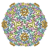
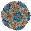
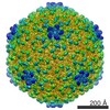
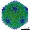
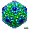
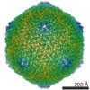
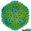
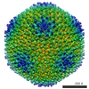
| Title | High resolution electron cryo-microscopy structure of the bacteriophage PR772 | ||||||||||||
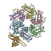
 Components Components |
| ||||||||||||
 Keywords Keywords | VIRUS / Phage / Tectiviridae / Membrane / double-barrel / helix turn helix / helix with a kink / beta-propeller / heteropentamer / penton | ||||||||||||
| Function / homology |  Function and homology information Function and homology information | ||||||||||||
| Biological species |  Enterobacteria phage PR772 (virus) Enterobacteria phage PR772 (virus) | ||||||||||||
| Method | ELECTRON MICROSCOPY / single particle reconstruction / cryo EM / Resolution: 2.75 Å | ||||||||||||
 Authors Authors | Narayana Reddy, H.K. / Svenda, M. | ||||||||||||
| Funding support |  Sweden, 3items Sweden, 3items
| ||||||||||||
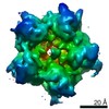
 Citation Citation | Journal: Elife / Year: 2019 Title: Electron cryo-microscopy of bacteriophage PR772 reveals the elusive vertex complex and the capsid architecture. Authors: Hemanth Kn Reddy / Marta Carroni / Janos Hajdu / Martin Svenda /  Abstract: Bacteriophage PR772, a member of the family, has a 70 nm diameter icosahedral protein capsid that encapsulates a lipid membrane, dsDNA, and various internal proteins. An icosahedrally averaged ...Bacteriophage PR772, a member of the family, has a 70 nm diameter icosahedral protein capsid that encapsulates a lipid membrane, dsDNA, and various internal proteins. An icosahedrally averaged CryoEM reconstruction of the wild-type virion and a localized reconstruction of the vertex region reveal the composition and the structure of the vertex complex along with new protein conformations that play a vital role in maintaining the capsid architecture of the virion. The overall resolution of the virion is 2.75 Å, while the resolution of the protein capsid is 2.3 Å. The conventional penta-symmetron formed by the capsomeres is replaced by a large vertex complex in the pseudo T = 25 capsid. All the vertices contain the host-recognition protein, P5; two of these vertices show the presence of the receptor-binding protein, P2. The 3D structure of the vertex complex shows interactions with the viral membrane, indicating a possible mechanism for viral infection. #1: Journal: Biorxiv / Year: 2019Title: Electron cryo-microscopy of Bacteriophage PR772 reveals the composition and structure of the elusive vertex complex and the capsid architecture Authors: Narayana Reddy, H.K. / Hajdu, J. / Carroni, M. / Svenda, M. | ||||||||||||
| History |
|
- Structure visualization
| Movie |
Movie viewer |
|---|---|
| Structure viewer | Molecule: MolmilJmol/JSmol |
- Downloads & links
-Download
| PDBx/mmCIF format | 6q5u.cif.gz | 898.4 KB | Display | PDBx/mmCIF format |
|---|---|---|---|---|
| PDB format | pdb6q5u.ent.gz | 752.3 KB | Display | PDB format |
| PDBx/mmJSON format | 6q5u.json.gz | Tree view | PDBx/mmJSON format | |
| Others |  Other downloads Other downloads |
-Validation report
| Arichive directory | https://data.pdbj.org/pub/pdb/validation_reports/q5/6q5uftp://data.pdbj.org/pub/pdb/validation_reports/q5/6q5u | HTTPS FTP |
|---|
-Related structure data
| Related structure data | 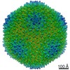 4461MC 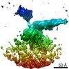 4462C M: map data used to model this data C: citing same article ( |
|---|---|
| Similar structure data |


-Links
 PDBj
PDBj

- Assembly
| Deposited unit | 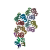
|
|---|---|
| 1 | x 60 x 12 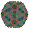
|
-Components
| #1: Protein | Mass: 43505.492 Da / Num. of mol.: 12 / Source method: isolated from a natural source / Source: (natural) Enterobacteria phage PR772 (virus) / References: UniProt: Q6EDX0#2: Protein | | Mass: 9275.559 Da / Num. of mol.: 1 / Source method: isolated from a natural source / Source: (natural) Enterobacteria phage PR772 (virus) / References: UniProt: Q6EDW3#3: Protein | | Mass: 12616.262 Da / Num. of mol.: 1 / Source method: isolated from a natural source / Source: (natural) Enterobacteria phage PR772 (virus) / References: UniProt: Q6EDW1#4: Protein | Mass: 34483.898 Da / Num. of mol.: 3 / Source method: isolated from a natural source / Source: (natural) Enterobacteria phage PR772 (virus) / References: UniProt: Q6EDX8#5: Protein | Mass: 13757.388 Da / Num. of mol.: 2 / Source method: isolated from a natural source / Source: (natural) Enterobacteria phage PR772 (virus) / References: UniProt: Q6EDY0 |
|---|
-Experimental details
-Experiment
| Experiment | Method: ELECTRON MICROSCOPY |
|---|---|
| EM experiment | Aggregation state: PARTICLE / 3D reconstruction method: single particle reconstruction |
- Sample preparation
| Component | Name: Bacteriophage PR772 / Type: COMPLEX / Details: Wild type / Entity ID: all / Source: NATURAL |
|---|---|
| Molecular weight | Value: 86 MDa / Experimental value: NO |
| Source (natural) | Organism: Enterobacteria phage PR772 (virus) |
| Details of virus | Empty: NO / Enveloped: NO / Isolate: SEROTYPE / Type: VIRION |
| Natural host | Organism: Escherichia coli / Strain: C-3000 |
| Virus shell | Diameter: 750 nm / Triangulation number (T number): 25 |
| Buffer solution | pH: 8 |
| Specimen | Conc.: 7 mg/ml / Embedding applied: NO / Shadowing applied: NO / Staining applied: NO / Vitrification applied: YES |
| Specimen support | Grid material: COPPER / Grid type: C-flat-2/2 |
| Vitrification | Instrument: FEI VITROBOT MARK IV / Cryogen name: ETHANE / Humidity: 100 % / Chamber temperature: 298.15 K |
- Electron microscopy imaging
| Experimental equipment |  Model: Titan Krios / Image courtesy: FEI Company |
|---|---|
| Microscopy | Model: FEI TITAN KRIOS |
| Electron gun | Electron source:  FIELD EMISSION GUN / Accelerating voltage: 300 kV / Illumination mode: FLOOD BEAM FIELD EMISSION GUN / Accelerating voltage: 300 kV / Illumination mode: FLOOD BEAM |
| Electron lens | Mode: BRIGHT FIELD / Nominal magnification: 130000 X / Nominal defocus max: 2600 nm / Nominal defocus min: 800 nm / Cs: 2.7 mm |
| Specimen holder | Cryogen: NITROGEN |
| Image recording | Electron dose: 40 e/Å2 / Film or detector model: GATAN K2 SUMMIT (4k x 4k) / Num. of grids imaged: 1 / Num. of real images: 3200 |
| Image scans | Movie frames/image: 40 / Used frames/image: 3-40 |
- Processing
| Software | Name: PHENIX / Version: 1.14_3260: / Classification: refinement | ||||||||||||||||||||||||
|---|---|---|---|---|---|---|---|---|---|---|---|---|---|---|---|---|---|---|---|---|---|---|---|---|---|
| EM software |
| ||||||||||||||||||||||||
| CTF correction | Type: PHASE FLIPPING AND AMPLITUDE CORRECTION | ||||||||||||||||||||||||
| Particle selection | Num. of particles selected: 56000 | ||||||||||||||||||||||||
| Symmetry | Point symmetry: I (icosahedral) | ||||||||||||||||||||||||
| 3D reconstruction | Resolution: 2.75 Å / Resolution method: FSC 0.143 CUT-OFF / Num. of particles: 46000 / Symmetry type: POINT | ||||||||||||||||||||||||
| Atomic model building | B value: 104.93 / Protocol: AB INITIO MODEL / Space: REAL / Target criteria: Cross-correlation coefficient | ||||||||||||||||||||||||
| Refinement | Highest resolution: 2.75 Å / Stereochemistry target values: CORRELATION COEFFCIENT | ||||||||||||||||||||||||
| Refine LS restraints |
|
