 Movie
Movie Controller


+ Open data

- Basic information
| Entry | Database: EMDB / ID: EMD-5947 | |||||||||
|---|---|---|---|---|---|---|---|---|---|---|
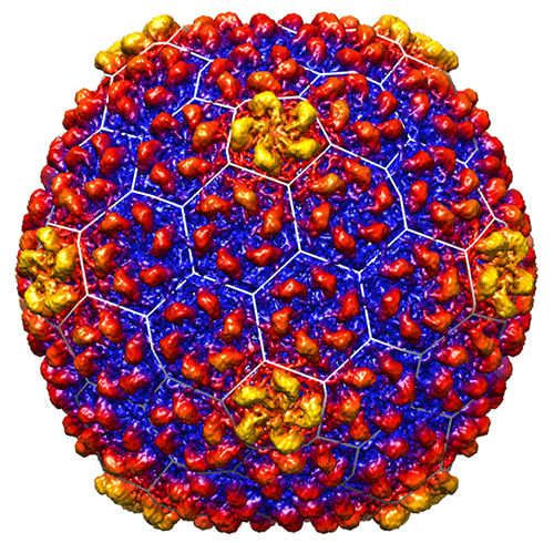
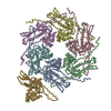
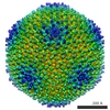
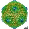
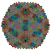
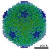
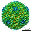
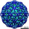
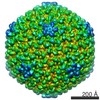
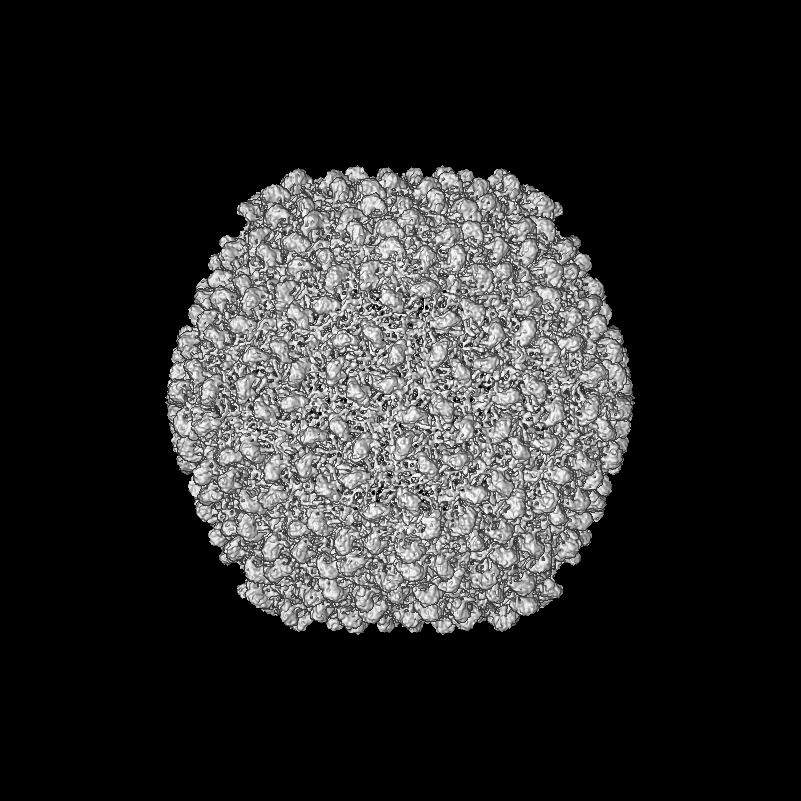
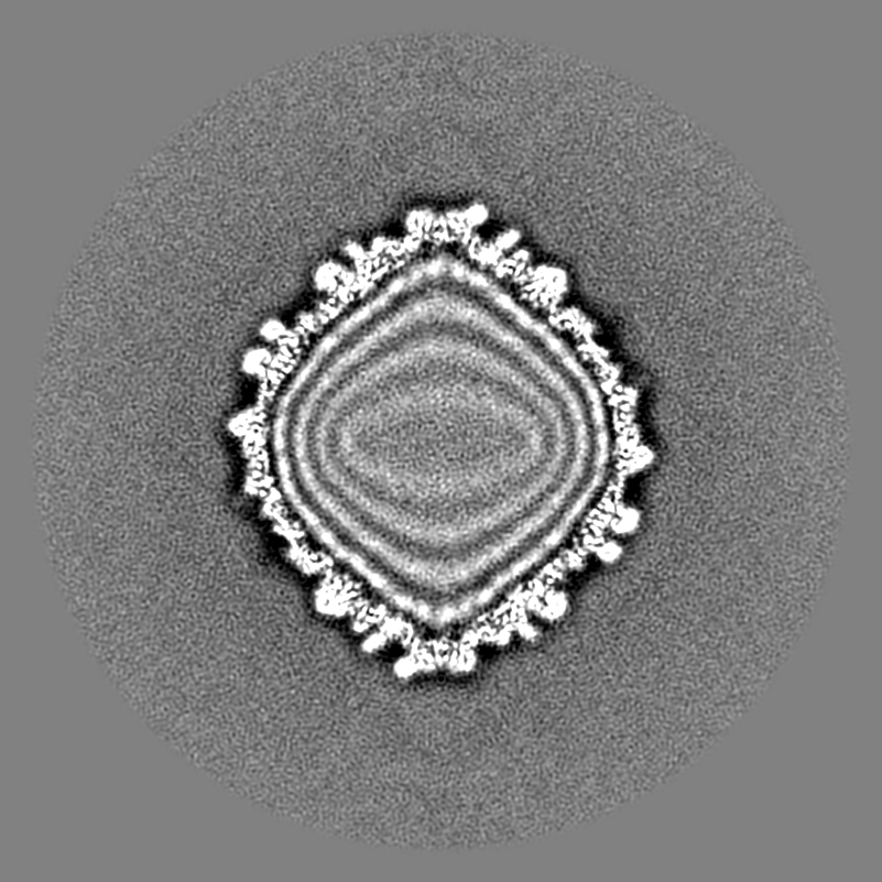
| Title | Bacteriophage CUS-3 capsid icosahedral reconstruction | |||||||||
 Map data Map data | Bacteriophage CUS-3 capsid icosahedral reconstruction | |||||||||
 Sample Sample |
| |||||||||
 Keywords Keywords | mature virion / capsid only / icosahedrally averaged | |||||||||
| Biological species |  Enterobacteria phage CUS-3 (virus) Enterobacteria phage CUS-3 (virus) | |||||||||
| Method | single particle reconstruction / cryo EM / Resolution: 6.8 Å | |||||||||
 Authors Authors | Parent KN / Tang J / Cardone G / Gilcrease EB / Janssen ME / Olson NH / Casjens SR / Baker TS | |||||||||
 Citation Citation | Journal: Virology / Year: 2014 Title: Three-dimensional reconstructions of the bacteriophage CUS-3 virion reveal a conserved coat protein I-domain but a distinct tailspike receptor-binding domain. Authors: Kristin N Parent / Jinghua Tang / Giovanni Cardone / Eddie B Gilcrease / Mandy E Janssen / Norman H Olson / Sherwood R Casjens / Timothy S Baker /  Abstract: CUS-3 is a short-tailed, dsDNA bacteriophage that infects serotype K1 Escherichia coli. We report icosahedrally averaged and asymmetric, three-dimensional, cryo-electron microscopic reconstructions ...CUS-3 is a short-tailed, dsDNA bacteriophage that infects serotype K1 Escherichia coli. We report icosahedrally averaged and asymmetric, three-dimensional, cryo-electron microscopic reconstructions of the CUS-3 virion. Its coat protein structure adopts the "HK97-fold" shared by other tailed phages and is quite similar to that in phages P22 and Sf6 despite only weak amino acid sequence similarity. In addition, these coat proteins share a unique extra external domain ("I-domain"), suggesting that the group of P22-like phages has evolved over a very long time period without acquiring a new coat protein gene from another phage group. On the other hand, the morphology of the CUS-3 tailspike differs significantly from that of P22 or Sf6, but is similar to the tailspike of phage K1F, a member of the extremely distantly related T7 group of phages. We conclude that CUS-3 obtained its tailspike gene from a distantly related phage quite recently. | |||||||||
| History |
|
- Structure visualization
| Movie |
 Movie viewer Movie viewer |
|---|---|
| Structure viewer | EM map: SurfViewMolmilJmol/JSmol |
| Supplemental images |
- Downloads & links
-EMDB archive
| Map data | emd_5947.map.gz | 602.6 MB | EMDB map data format | |
|---|---|---|---|---|
| Header (meta data) | emd-5947-v30.xmlemd-5947.xml | 10.3 KB 10.3 KB | Display Display | EMDB header |
| Images |  emd_5947.png emd_5947.png | 147.5 KB | ||
| Archive directory |  http://ftp.pdbj.org/pub/emdb/structures/EMD-5947ftp://ftp.pdbj.org/pub/emdb/structures/EMD-5947 http://ftp.pdbj.org/pub/emdb/structures/EMD-5947ftp://ftp.pdbj.org/pub/emdb/structures/EMD-5947 | HTTPS FTP |
-Related structure data
-Links
| EMDB pages | EMDB (EBI/PDBe) / EMDataResource |
|---|
-Map
| File | Download / File: emd_5947.map.gz / Format: CCP4 / Size: 1.9 GB / Type: IMAGE STORED AS FLOATING POINT NUMBER (4 BYTES) | ||||||||||||||||||||||||||||||||||||||||||||||||||||||||||||||||||||
|---|---|---|---|---|---|---|---|---|---|---|---|---|---|---|---|---|---|---|---|---|---|---|---|---|---|---|---|---|---|---|---|---|---|---|---|---|---|---|---|---|---|---|---|---|---|---|---|---|---|---|---|---|---|---|---|---|---|---|---|---|---|---|---|---|---|---|---|---|---|
| Annotation | Bacteriophage CUS-3 capsid icosahedral reconstruction | ||||||||||||||||||||||||||||||||||||||||||||||||||||||||||||||||||||
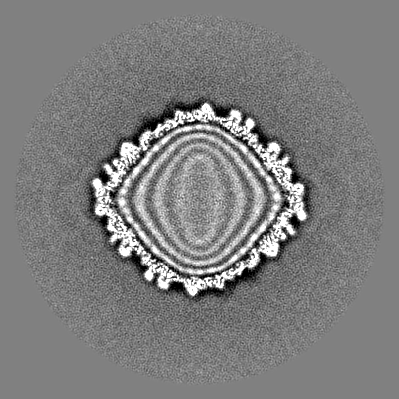
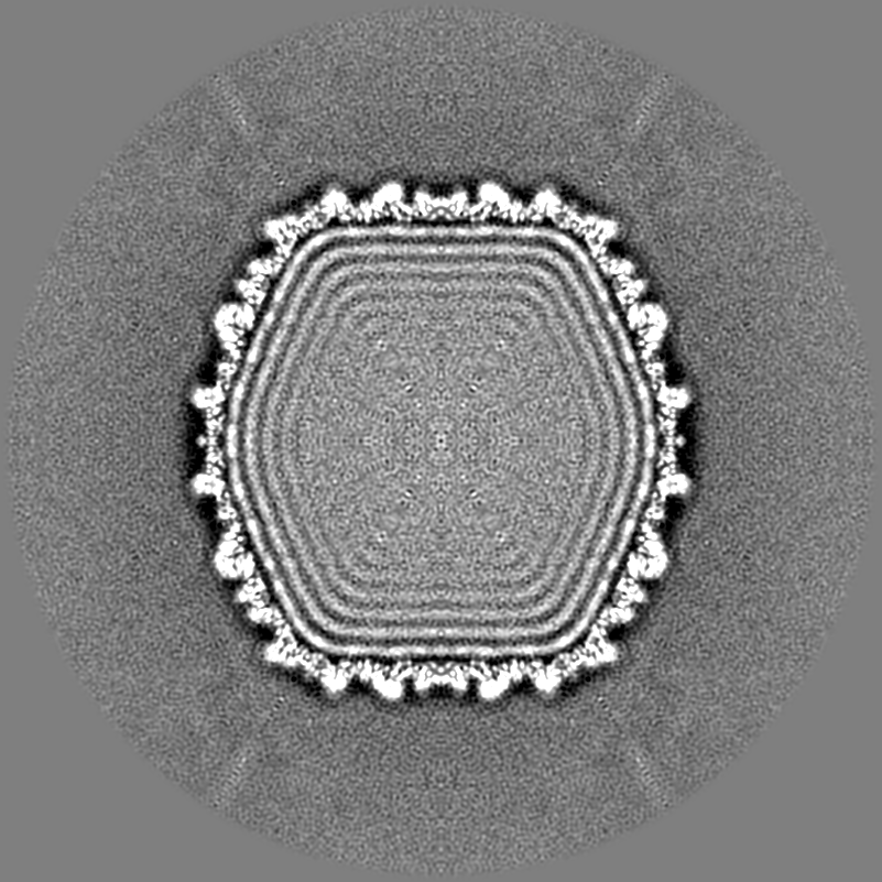
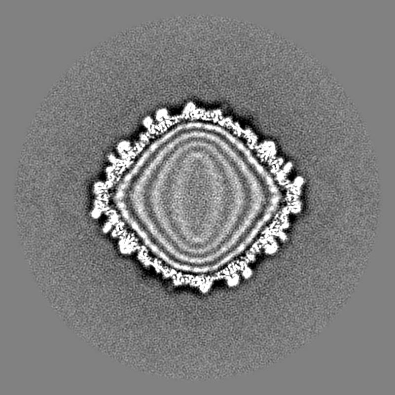
| Projections & slices | Image control
Images are generated by Spider. | ||||||||||||||||||||||||||||||||||||||||||||||||||||||||||||||||||||
| Voxel size | X=Y=Z: 1.35 Å | ||||||||||||||||||||||||||||||||||||||||||||||||||||||||||||||||||||
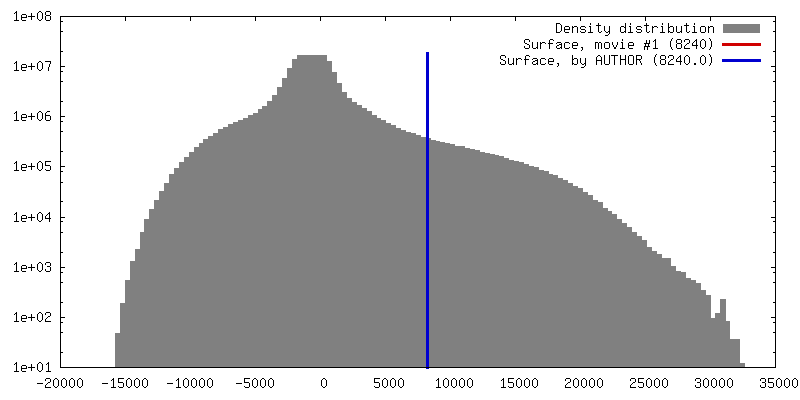
| Density |
| ||||||||||||||||||||||||||||||||||||||||||||||||||||||||||||||||||||
| Symmetry | Space group: 1 | ||||||||||||||||||||||||||||||||||||||||||||||||||||||||||||||||||||
| Details | EMDB XML:
CCP4 map header:
| ||||||||||||||||||||||||||||||||||||||||||||||||||||||||||||||||||||
 Z (Sec.)
 Y (Row.)
 X (Col.)


-Supplemental data
- Sample components
-Entire : CUS-3 virion, icosahedrally averaged
| Entire | Name: CUS-3 virion, icosahedrally averaged |
|---|---|
| Components |
|
-Supramolecule #1000: CUS-3 virion, icosahedrally averaged
| Supramolecule | Name: CUS-3 virion, icosahedrally averaged / type: sample / ID: 1000 / Oligomeric state: icosahedral / Number unique components: 1 |
|---|
-Supramolecule #1: Enterobacteria phage CUS-3
| Supramolecule | Name: Enterobacteria phage CUS-3 / type: virus / ID: 1 / NCBI-ID: 539221 / Sci species name: Enterobacteria phage CUS-3 / Database: NCBI / Virus type: VIRION / Virus isolate: SPECIES / Virus enveloped: No / Virus empty: No |
|---|---|
| Host (natural) | Organism:  |
| Virus shell | Shell ID: 1 / Name: capsid / Diameter: 690 Å / T number (triangulation number): 7 |
-Experimental details
-Structure determination
| Method | cryo EM |
|---|---|
 Processing Processing | single particle reconstruction |
| Aggregation state | particle |
-Sample preparation
| Concentration | 10 mg/mL |
|---|---|
| Buffer | pH: 7.6 / Details: 10 mM Tris, 10 mM MgCl2 |
| Grid | Details: 400 mesh R2/2 Quantifoil, glow discharged |
| Vitrification | Cryogen name: ETHANE / Chamber humidity: 100 % / Chamber temperature: 90 K / Instrument: HOMEMADE PLUNGER / Method: Blot for 5 sec before plunging. |
- Electron microscopy
| Microscope | FEI POLARA 300 |
|---|---|
| Temperature | Min: 89 K / Max: 91 K / Average: 90 K |
| Alignment procedure | Legacy - Astigmatism: Objective lens astigmatism was corrected at the magnification used to collect data. |
| Date | Sep 1, 2013 |
| Image recording | Category: CCD / Film or detector model: DIRECT ELECTRON DE-12 (4k x 3k) / Digitization - Sampling interval: 6 µm / Number real images: 419 / Average electron dose: 23 e/Å2 Details: The data were collected on the DE12 camera under control of the automated acquisition software, LEGINON. |
| Electron beam | Acceleration voltage: 200 kV / Electron source:  FIELD EMISSION GUN FIELD EMISSION GUN |
| Electron optics | Illumination mode: FLOOD BEAM / Imaging mode: BRIGHT FIELD / Cs: 2.3 mm / Nominal defocus max: 4.08 µm / Nominal defocus min: 0.6 µm / Nominal magnification: 31000 |
| Sample stage | Specimen holder model: OTHER |
| Experimental equipment |  Model: Tecnai Polara / Image courtesy: FEI Company |
-Image processing
| Details | Auto3dem was used for refinement. |
|---|---|
| CTF correction | Details: each micrograph |
| Final reconstruction | Algorithm: OTHER / Resolution.type: BY AUTHOR / Resolution: 6.8 Å / Resolution method: OTHER / Software - Name: Auto3dem, RobEM, autopp Details: 28 frames total were collected for each image (35 e-/A2 total dose). Only 15 were used in the final reconstruction (18e-/A2 dose). Number images used: 7766 |
