National Institutes of Health/National Institute of General Medical Sciences (NIH/NIGMS)
F32GM108310
United States
Citation
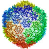
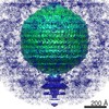
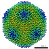
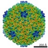
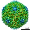
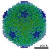
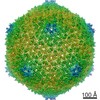
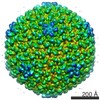
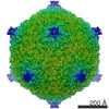
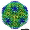
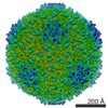
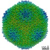
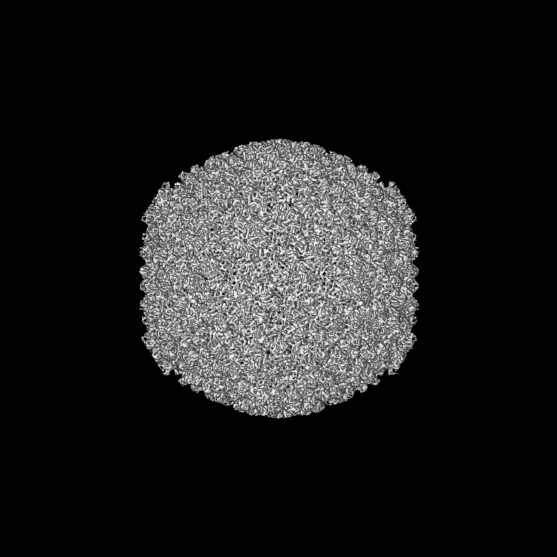
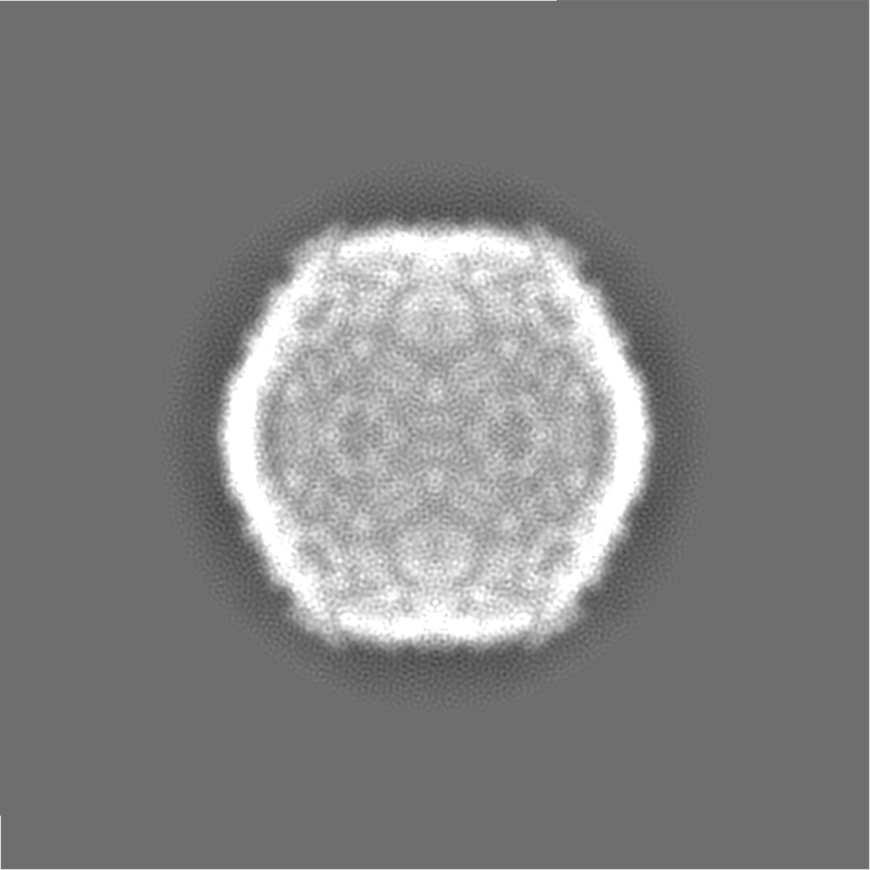
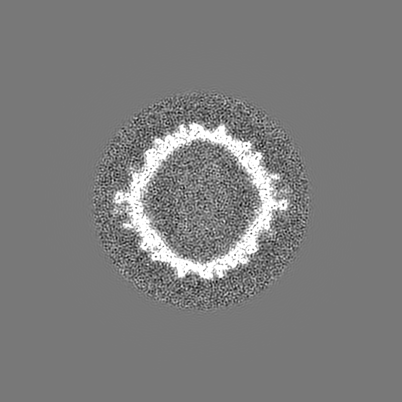
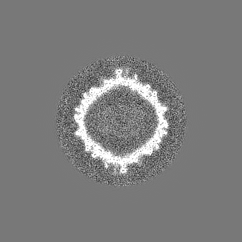
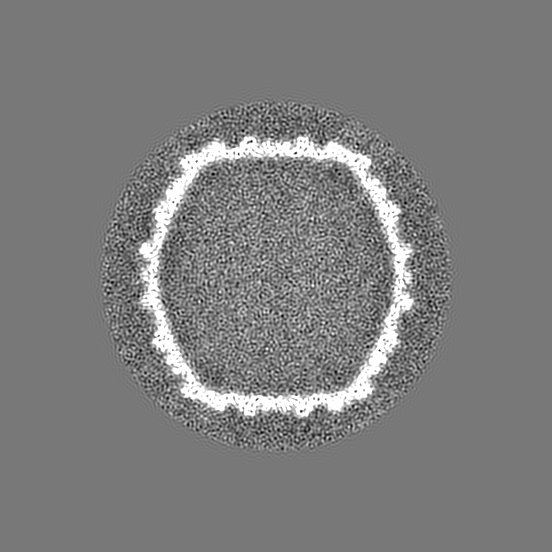
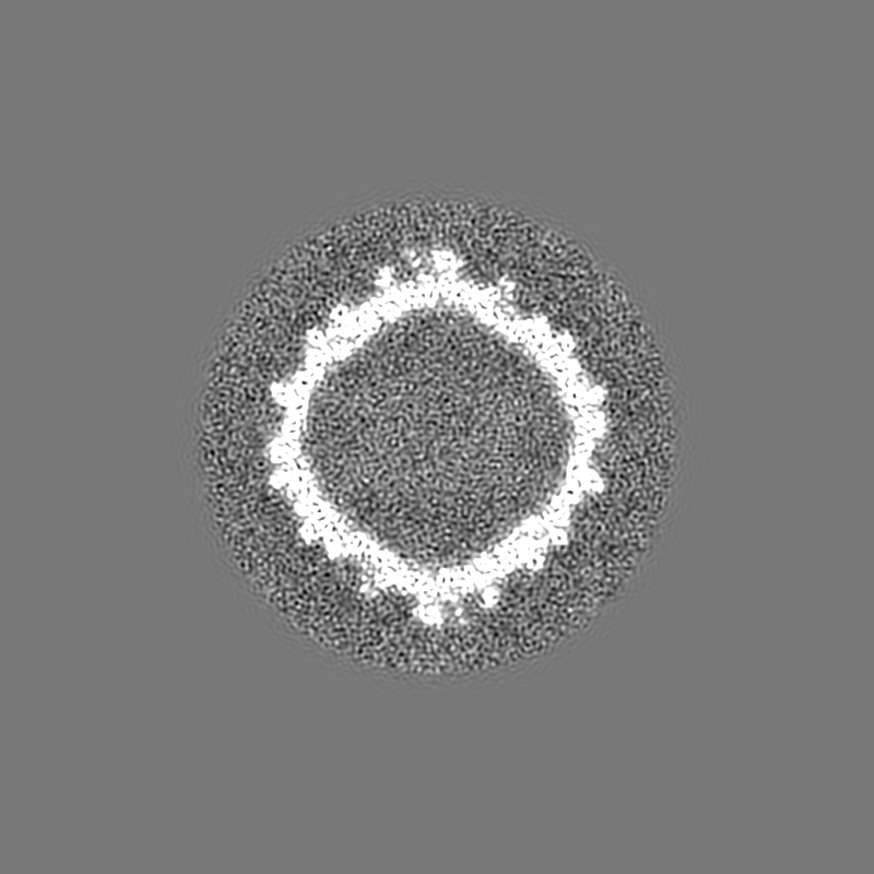
Journal: Biophys J / Year: 2018 Title: Cryo-EM Elucidation of the Structure of Bacteriophage P22 Virions after Genome Release. Authors: Reginald McNulty / Giovanni Cardone / Eddie B Gilcrease / Timothy S Baker / Sherwood R Casjens / John E Johnson / Abstract: Genome ejection proteins are required to facilitate transport of bacteriophage P22 double-stranded DNA safely through membranes of Salmonella. The structures and locations of all proteins in the ...Genome ejection proteins are required to facilitate transport of bacteriophage P22 double-stranded DNA safely through membranes of Salmonella. The structures and locations of all proteins in the context of the mature virion are known, with the exception of three ejection proteins. Furthermore, the changes that occur to the proteins residing in the mature virion upon DNA release are not fully understood. We used cryogenic electron microscopy to obtain what is, to our knowledge, the first asymmetric reconstruction of mature bacteriophage P22 after double-stranded DNA has been extruded from the capsid-a state representative of one step during viral infection. Results of icosahedral and asymmetric reconstructions at estimated resolutions of 7.8 and 12.5 Å resolutions, respectively, are presented. The reconstruction shows tube-like protein density extending from the center of the tail assembly. The portal protein does not revert to the more contracted, procapsid state, but instead maintains an extended and splayed barrel structure. These structural details contribute to our understanding of the molecular mechanism of P22 phage infection and also set the foundation for future exploitation serving engineering purposes.
History
Deposition
Dec 23, 2017
-
Header (metadata) release
Jan 24, 2018
-
Map release
Apr 18, 2018
-
Update
Jan 29, 2020
-
Current status
Jan 29, 2020
Processing site: RCSB / Status: Released
-
Structure visualization
Movie
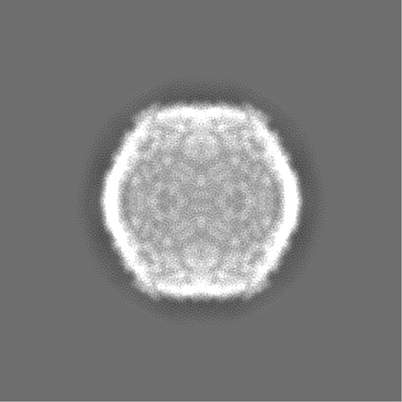
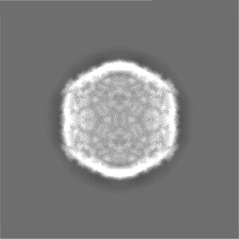
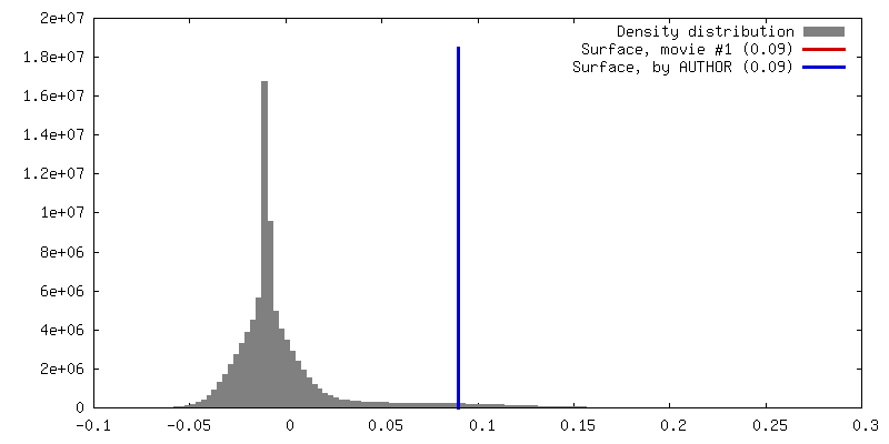
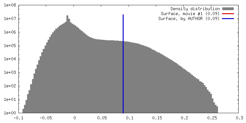
Surface view with section colored by density value
In the structure databanks used in Yorodumi, some data are registered as the other names, "COVID-19 virus" and "2019-nCoV". Here are the details of the virus and the list of structure data.
Jan 31, 2019. EMDB accession codes are about to change! (news from PDBe EMDB page)
EMDB accession codes are about to change! (news from PDBe EMDB page)
The allocation of 4 digits for EMDB accession codes will soon come to an end. Whilst these codes will remain in use, new EMDB accession codes will include an additional digit and will expand incrementally as the available range of codes is exhausted. The current 4-digit format prefixed with “EMD-” (i.e. EMD-XXXX) will advance to a 5-digit format (i.e. EMD-XXXXX), and so on. It is currently estimated that the 4-digit codes will be depleted around Spring 2019, at which point the 5-digit format will come into force.
The EM Navigator/Yorodumi systems omit the EMD- prefix.
Related info.:Q: What is EMD? / ID/Accession-code notation in Yorodumi/EM Navigator
Yorodumi is a browser for structure data from EMDB, PDB, SASBDB, etc.
This page is also the successor to EM Navigator detail page, and also detail information page/front-end page for Omokage search.
The word "yorodu" (or yorozu) is an old Japanese word meaning "ten thousand". "mi" (miru) is to see.
Related info.:EMDB / PDB / SASBDB / Comparison of 3 databanks / Yorodumi Search / Aug 31, 2016. New EM Navigator & Yorodumi / Yorodumi Papers / Jmol/JSmol / Function and homology information / Changes in new EM Navigator and Yorodumi
 Movie
Movie Controller


 Open data
 Basic information
 Map data
 Sample
 Enterobacteria phage P22 (virus)
 Authors
 United States, 1 items
  Citation
 Structure visualization
 Movie viewer
 Downloads & links
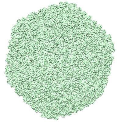 emd_7315.png
 http://ftp.pdbj.org/pub/emdb/structures/EMD-7315
 Z (Sec.)
 Y (Row.)
 X (Col.)


 Sample components
 Salmonella enterica (bacteria)
 Processing
 Electron microscopy
 FIELD EMISSION GUN