 ムービー
ムービー コントローラー


+ データを開く

- 基本情報
基本情報
| 登録情報 | データベース: PDB / ID: 6q5u | ||||||||||||
|---|---|---|---|---|---|---|---|---|---|---|---|---|---|
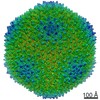
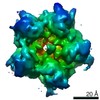
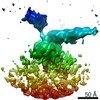
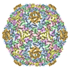
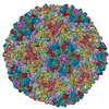
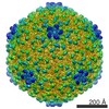
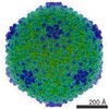
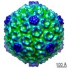
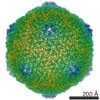
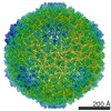
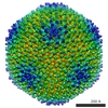
| タイトル | High resolution electron cryo-microscopy structure of the bacteriophage PR772 | ||||||||||||
 要素 要素 |
| ||||||||||||
 キーワード キーワード |  VIRUS (ウイルス) / Phage (ファージ) / Tectiviridae / Membrane (生体膜) / double-barrel / helix turn helix / helix with a kink / beta-propeller (Βプロペラドメイン) / heteropentamer / penton VIRUS (ウイルス) / Phage (ファージ) / Tectiviridae / Membrane (生体膜) / double-barrel / helix turn helix / helix with a kink / beta-propeller (Βプロペラドメイン) / heteropentamer / penton | ||||||||||||
| 機能・相同性 |  機能・相同性情報 機能・相同性情報 | ||||||||||||
| 生物種 |  Enterobacteria phage PR772 (ファージ) Enterobacteria phage PR772 (ファージ) | ||||||||||||
| 手法 | 電子顕微鏡法 / 単粒子再構成法 / クライオ電子顕微鏡法 / 解像度: 2.75 Å | ||||||||||||
 データ登録者 データ登録者 | Narayana Reddy, H.K. / Svenda, M. | ||||||||||||
| 資金援助 |  スウェーデン, 3件 スウェーデン, 3件
| ||||||||||||
 引用 引用 | ジャーナル: Elife / 年: 2019 タイトル: Electron cryo-microscopy of bacteriophage PR772 reveals the elusive vertex complex and the capsid architecture. 著者: Hemanth Kn Reddy / Marta Carroni / Janos Hajdu / Martin Svenda /  要旨: Bacteriophage PR772, a member of the family, has a 70 nm diameter icosahedral protein capsid that encapsulates a lipid membrane, dsDNA, and various internal proteins. An icosahedrally averaged ...Bacteriophage PR772, a member of the family, has a 70 nm diameter icosahedral protein capsid that encapsulates a lipid membrane, dsDNA, and various internal proteins. An icosahedrally averaged CryoEM reconstruction of the wild-type virion and a localized reconstruction of the vertex region reveal the composition and the structure of the vertex complex along with new protein conformations that play a vital role in maintaining the capsid architecture of the virion. The overall resolution of the virion is 2.75 Å, while the resolution of the protein capsid is 2.3 Å. The conventional penta-symmetron formed by the capsomeres is replaced by a large vertex complex in the pseudo T = 25 capsid. All the vertices contain the host-recognition protein, P5; two of these vertices show the presence of the receptor-binding protein, P2. The 3D structure of the vertex complex shows interactions with the viral membrane, indicating a possible mechanism for viral infection. #1: ジャーナル: Biorxiv / 年: 2019タイトル: Electron cryo-microscopy of Bacteriophage PR772 reveals the composition and structure of the elusive vertex complex and the capsid architecture 著者: Narayana Reddy, H.K. / Hajdu, J. / Carroni, M. / Svenda, M. | ||||||||||||
| 履歴 |
|
- 構造の表示
構造の表示
| ムービー |
ムービービューア |
|---|---|
| 構造ビューア | 分子: MolmilJmol/JSmol |
- ダウンロードとリンク
-ダウンロード
| PDBx/mmCIF形式 | 6q5u.cif.gz | 893.7 KB | 表示 | PDBx/mmCIF形式 |
|---|---|---|---|---|
| PDB形式 | pdb6q5u.ent.gz | 765.7 KB | 表示 | PDB形式 |
| PDBx/mmJSON形式 | 6q5u.json.gz | ツリー表示 | PDBx/mmJSON形式 | |
| その他 |  その他のダウンロード その他のダウンロード |
-検証レポート
| アーカイブディレクトリ | https://data.pdbj.org/pub/pdb/validation_reports/q5/6q5uftp://data.pdbj.org/pub/pdb/validation_reports/q5/6q5u | HTTPS FTP |
|---|
-関連構造データ


-リンク
 PDBj
PDBj

- 集合体
集合体
| 登録構造単位 | 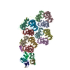
|
|---|---|
| 1 | x 60 x 12 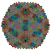
|
-要素
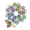
| #1: タンパク質 | 分子量: 43505.492 Da / 分子数: 12 / 由来タイプ: 天然 / 由来: (天然) Enterobacteria phage PR772 (ファージ) / 参照: UniProt: Q6EDX0#2: タンパク質 | | 分子量: 9275.559 Da / 分子数: 1 / 由来タイプ: 天然 / 由来: (天然) Enterobacteria phage PR772 (ファージ) / 参照: UniProt: Q6EDW3#3: タンパク質 | | 分子量: 12616.262 Da / 分子数: 1 / 由来タイプ: 天然 / 由来: (天然) Enterobacteria phage PR772 (ファージ) / 参照: UniProt: Q6EDW1#4: タンパク質 | スパイクタンパク質分子量: 34483.898 Da / 分子数: 3 / 由来タイプ: 天然 / 由来: (天然) Enterobacteria phage PR772 (ファージ) / 参照: UniProt: Q6EDX8#5: タンパク質 | 分子量: 13757.388 Da / 分子数: 2 / 由来タイプ: 天然 / 由来: (天然) Enterobacteria phage PR772 (ファージ) / 参照: UniProt: Q6EDY0 |
|---|
-実験情報
-実験
| 実験 | 手法: 電子顕微鏡法 |
|---|---|
| EM実験 | 試料の集合状態: PARTICLE / 3次元再構成法: 単粒子再構成法 |
- 試料調製
試料調製
| 構成要素 | 名称: Bacteriophage PR772 / タイプ: COMPLEX / 詳細: Wild type / Entity ID: all / 由来: NATURAL |
|---|---|
| 分子量 | 値: 86 MDa / 実験値: NO |
| 由来(天然) | 生物種: Enterobacteria phage PR772 (ファージ) |
| ウイルスについての詳細 | 中空か: NO / エンベロープを持つか: NO / 単離: SEROTYPE / タイプ: VIRION |
| 天然宿主 | 生物種: Escherichia coli / 株: C-3000 |
| ウイルス殻 | 直径: 750 nm / 三角数 (T数): 25 |
| 緩衝液 | pH: 8 |
| 試料 | 濃度: 7 mg/ml / 包埋: NO / シャドウイング: NO / 染色: NO / 凍結: YES |
| 試料支持 | グリッドの材料: COPPER / グリッドのタイプ: C-flat-2/2 |
| 急速凍結 | 装置: FEI VITROBOT MARK IV / 凍結剤: ETHANE / 湿度: 100 % / 凍結前の試料温度: 298.15 K |
- 電子顕微鏡撮影
| 実験機器 |  モデル: Titan Krios / 画像提供: FEI Company |
|---|---|
| 顕微鏡 | モデル: FEI TITAN KRIOS |
| 電子銃 | 電子線源: FIELD EMISSION GUN / 加速電圧: 300 kV / 照射モード: FLOOD BEAM |
| 電子レンズ | モード: BRIGHT FIELDBright-field microscopy / 倍率(公称値): 130000 X / 最大 デフォーカス(公称値): 2600 nm / 最小 デフォーカス(公称値): 800 nm / Cs: 2.7 mm |
| 試料ホルダ | 凍結剤: NITROGEN |
| 撮影 | 電子線照射量: 40 e/Å2 フィルム・検出器のモデル: GATAN K2 SUMMIT (4k x 4k) 撮影したグリッド数: 1 / 実像数: 3200 |
| 画像スキャン | 動画フレーム数/画像: 40 / 利用したフレーム数/画像: 3-40 |
- 解析
解析
| ソフトウェア | 名称: PHENIX / バージョン: 1.14_3260: / 分類: 精密化 | ||||||||||||||||||||||||
|---|---|---|---|---|---|---|---|---|---|---|---|---|---|---|---|---|---|---|---|---|---|---|---|---|---|
| EMソフトウェア |
| ||||||||||||||||||||||||
| CTF補正 | タイプ: PHASE FLIPPING AND AMPLITUDE CORRECTION | ||||||||||||||||||||||||
| 粒子像の選択 | 選択した粒子像数: 56000 | ||||||||||||||||||||||||
| 対称性 | 点対称性: I (正20面体型対称) | ||||||||||||||||||||||||
| 3次元再構成 | 解像度: 2.75 Å / 解像度の算出法: FSC 0.143 CUT-OFF / 粒子像の数: 46000 / 対称性のタイプ: POINT | ||||||||||||||||||||||||
| 原子モデル構築 | B value: 104.93 / プロトコル: AB INITIO MODEL / 空間: REAL / Target criteria: Cross-correlation coefficient | ||||||||||||||||||||||||
| 精密化 | 最高解像度: 2.75 Å / 立体化学のターゲット値: CORRELATION COEFFCIENT | ||||||||||||||||||||||||
| 拘束条件 |
|
