 Movie
Movie Controller

[English] 日本語
 Yorodumi
- EMDB-10238: Focused Classification of the vertex region of Bacteriophage PR77... -
+ Open data

- Basic information
| Entry | Database: EMDB / ID: EMD-10238 | ||||||||||||
|---|---|---|---|---|---|---|---|---|---|---|---|---|---|
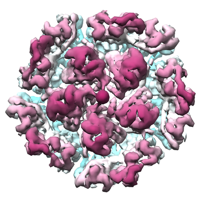
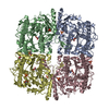
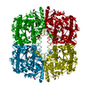
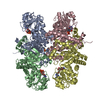
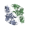
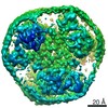
| Title | Focused Classification of the vertex region of Bacteriophage PR772 showing the heteropentameric penton | ||||||||||||
 Map data Map data | Focused Classification of the vertex region of Bacteriophage PR772 showing the heteropentameric penton | ||||||||||||
 Sample Sample |
| ||||||||||||
| Biological species |  Enterobacteria phage PR772 (virus) Enterobacteria phage PR772 (virus) | ||||||||||||
| Method | single particle reconstruction / cryo EM / Resolution: 4.25 Å | ||||||||||||
 Authors Authors | Narayana Reddy HK / Svenda M | ||||||||||||
| Funding support |  Sweden, 3 items Sweden, 3 items
| ||||||||||||
 Citation Citation | Journal: Elife / Year: 2019 Title: Electron cryo-microscopy of bacteriophage PR772 reveals the elusive vertex complex and the capsid architecture. Authors: Hemanth Kn Reddy / Marta Carroni / Janos Hajdu / Martin Svenda /  Abstract: Bacteriophage PR772, a member of the family, has a 70 nm diameter icosahedral protein capsid that encapsulates a lipid membrane, dsDNA, and various internal proteins. An icosahedrally averaged ...Bacteriophage PR772, a member of the family, has a 70 nm diameter icosahedral protein capsid that encapsulates a lipid membrane, dsDNA, and various internal proteins. An icosahedrally averaged CryoEM reconstruction of the wild-type virion and a localized reconstruction of the vertex region reveal the composition and the structure of the vertex complex along with new protein conformations that play a vital role in maintaining the capsid architecture of the virion. The overall resolution of the virion is 2.75 Å, while the resolution of the protein capsid is 2.3 Å. The conventional penta-symmetron formed by the capsomeres is replaced by a large vertex complex in the pseudo T = 25 capsid. All the vertices contain the host-recognition protein, P5; two of these vertices show the presence of the receptor-binding protein, P2. The 3D structure of the vertex complex shows interactions with the viral membrane, indicating a possible mechanism for viral infection. | ||||||||||||
| History |
|
- Structure visualization
| Movie |
 Movie viewer Movie viewer |
|---|---|
| Structure viewer | EM map: SurfViewMolmilJmol/JSmol |
| Supplemental images |
- Downloads & links
-EMDB archive
| Map data | emd_10238.map.gz | 5.9 MB | EMDB map data format | |
|---|---|---|---|---|
| Header (meta data) | emd-10238-v30.xmlemd-10238.xml | 12.2 KB 12.2 KB | Display Display | EMDB header |
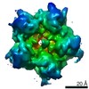
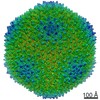
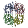
| Images |  emd_10238.png emd_10238.png | 263.5 KB | ||
| Archive directory |  http://ftp.pdbj.org/pub/emdb/structures/EMD-10238ftp://ftp.pdbj.org/pub/emdb/structures/EMD-10238 http://ftp.pdbj.org/pub/emdb/structures/EMD-10238ftp://ftp.pdbj.org/pub/emdb/structures/EMD-10238 | HTTPS FTP |
-Related structure data


-Links
| EMDB pages | EMDB (EBI/PDBe) / EMDataResource |
|---|
-Map
| File | Download / File: emd_10238.map.gz / Format: CCP4 / Size: 307.5 MB / Type: IMAGE STORED AS FLOATING POINT NUMBER (4 BYTES) | ||||||||||||||||||||||||||||||||||||||||||||||||||||||||||||
|---|---|---|---|---|---|---|---|---|---|---|---|---|---|---|---|---|---|---|---|---|---|---|---|---|---|---|---|---|---|---|---|---|---|---|---|---|---|---|---|---|---|---|---|---|---|---|---|---|---|---|---|---|---|---|---|---|---|---|---|---|---|
| Annotation | Focused Classification of the vertex region of Bacteriophage PR772 showing the heteropentameric penton | ||||||||||||||||||||||||||||||||||||||||||||||||||||||||||||
| Projections & slices | Image control
Images are generated by Spider. | ||||||||||||||||||||||||||||||||||||||||||||||||||||||||||||
| Voxel size | X=Y=Z: 2.12 Å | ||||||||||||||||||||||||||||||||||||||||||||||||||||||||||||
| Density |
| ||||||||||||||||||||||||||||||||||||||||||||||||||||||||||||
| Symmetry | Space group: 1 | ||||||||||||||||||||||||||||||||||||||||||||||||||||||||||||
| Details | EMDB XML:
CCP4 map header:
| ||||||||||||||||||||||||||||||||||||||||||||||||||||||||||||
 Z (Sec.)
 Y (Row.)
 X (Col.)


-Supplemental data
- Sample components
-Entire : Bacteriophage PR772
| Entire | Name: Bacteriophage PR772 |
|---|---|
| Components |
|
-Supramolecule #1: Bacteriophage PR772
| Supramolecule | Name: Bacteriophage PR772 / type: complex / ID: 1 / Parent: 0 / Macromolecule list: #1-#5 / Details: Wild type |
|---|---|
| Source (natural) | Organism: Enterobacteria phage PR772 (virus) |
| Molecular weight | Theoretical: 100 KDa |
-Experimental details
-Structure determination
| Method | cryo EM |
|---|---|
 Processing Processing | single particle reconstruction |
| Aggregation state | particle |
-Sample preparation
| Concentration | 7 mg/mL |
|---|---|
| Buffer | pH: 8 |
| Vitrification | Cryogen name: ETHANE / Chamber humidity: 100 % / Chamber temperature: 298.15 K / Instrument: FEI VITROBOT MARK IV |
- Electron microscopy
| Microscope | FEI TITAN KRIOS |
|---|---|
| Image recording | Film or detector model: GATAN K2 SUMMIT (4k x 4k) / Digitization - Frames/image: 3-40 / Number grids imaged: 1 / Number real images: 3200 / Average electron dose: 40.0 e/Å2 |
| Electron beam | Acceleration voltage: 300 kV / Electron source:  FIELD EMISSION GUN FIELD EMISSION GUN |
| Electron optics | Illumination mode: FLOOD BEAM / Imaging mode: BRIGHT FIELD / Cs: 2.7 mm / Nominal defocus max: 2.6 µm / Nominal defocus min: 0.8 µm / Nominal magnification: 130000 |
| Sample stage | Cooling holder cryogen: NITROGEN |
| Experimental equipment |  Model: Titan Krios / Image courtesy: FEI Company |
+Image processing
-Atomic model buiding 1
| Software | Name: Coot (ver. 0.8.9.1) |
|---|---|
| Refinement | Space: REAL / Protocol: AB INITIO MODEL / Overall B value: 104.93 / Target criteria: Cross-correlation coefficient |
