 Movie
Movie Controller

[English] 日本語
 Yorodumi
- EMDB-5142: Ab initio reconstruction of the avian polyomavirus via the asymme... -
+ Open data

- Basic information
| Entry | Database: EMDB / ID: EMD-5142 | |||||||||
|---|---|---|---|---|---|---|---|---|---|---|
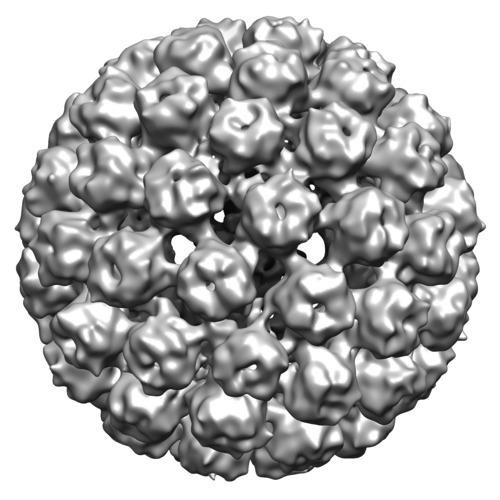
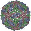
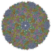
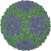
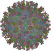
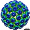
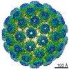
| Title | Ab initio reconstruction of the avian polyomavirus via the asymmetric random-model method | |||||||||
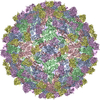
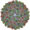
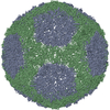
 Map data Map data | The avian polyomavirus | |||||||||
 Sample Sample |
| |||||||||
 Keywords Keywords | random-model method / ab initio reconstruction / avian polyomavirus | |||||||||
| Biological species |  Avian polyomavirus Avian polyomavirus | |||||||||
| Method | single particle reconstruction / cryo EM / Resolution: 35.0 Å | |||||||||
 Authors Authors | Sanz E / Stewart AB / Belnap DM | |||||||||
 Citation Citation | Journal: J Struct Biol / Year: 2010 Title: The random-model method enables ab initio 3D reconstruction of asymmetric particles and determination of particle symmetry. Authors: Eduardo Sanz-García / Aaron B Stewart / David M Belnap /  Abstract: Model-based, 3D reconstruction techniques depend on reliable starting models. We present an extension of the random-model method (RMM) that allows the ab initio generation of suitable starting models ...Model-based, 3D reconstruction techniques depend on reliable starting models. We present an extension of the random-model method (RMM) that allows the ab initio generation of suitable starting models directly from un-averaged, experimental images of asymmetric or symmetric particles. Therefore, the asymmetric RMM can also be used to determine point-group symmetry. The procedure is facilitated by the use of (a) variable angular step-sizes during iterative origin and orientation searches, (b) high numbers of particle images, and (c) highly defocused images. The method is inhibited by mixed-handedness orientation assignments and by particles with inconspicuous features. For symmetric particles, symmetric RMMs can overcome these deficiencies. | |||||||||
| History |
|
- Structure visualization
| Movie |
 Movie viewer Movie viewer |
|---|---|
| Structure viewer | EM map: SurfViewMolmilJmol/JSmol |
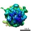
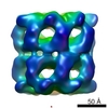
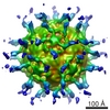
| Supplemental images |

- Downloads & links
-EMDB archive
| Map data | emd_5142.map.gz | 91.5 MB | EMDB map data format | |
|---|---|---|---|---|
| Header (meta data) | emd-5142-v30.xmlemd-5142.xml | 8.1 KB 8.1 KB | Display Display | EMDB header |
| Images |  400_5142.gif 400_5142.gif 80_5142.gifemd_5142_1.tif 80_5142.gifemd_5142_1.tif | 77.5 KB 6.3 KB 489.9 KB | ||
| Archive directory |  http://ftp.pdbj.org/pub/emdb/structures/EMD-5142ftp://ftp.pdbj.org/pub/emdb/structures/EMD-5142 http://ftp.pdbj.org/pub/emdb/structures/EMD-5142ftp://ftp.pdbj.org/pub/emdb/structures/EMD-5142 | HTTPS FTP |
-Related structure data
-Links
| EMDB pages | EMDB (EBI/PDBe) / EMDataResource |
|---|
-Map
| File | Download / File: emd_5142.map.gz / Format: CCP4 / Size: 184.1 MB / Type: IMAGE STORED AS FLOATING POINT NUMBER (4 BYTES) | ||||||||||||||||||||||||||||||||||||||||||||||||||||||||||||||||||||
|---|---|---|---|---|---|---|---|---|---|---|---|---|---|---|---|---|---|---|---|---|---|---|---|---|---|---|---|---|---|---|---|---|---|---|---|---|---|---|---|---|---|---|---|---|---|---|---|---|---|---|---|---|---|---|---|---|---|---|---|---|---|---|---|---|---|---|---|---|---|
| Annotation | The avian polyomavirus | ||||||||||||||||||||||||||||||||||||||||||||||||||||||||||||||||||||
| Projections & slices | Image control
Images are generated by Spider. | ||||||||||||||||||||||||||||||||||||||||||||||||||||||||||||||||||||
| Voxel size | X=Y=Z: 1.63 Å | ||||||||||||||||||||||||||||||||||||||||||||||||||||||||||||||||||||
| Density |
| ||||||||||||||||||||||||||||||||||||||||||||||||||||||||||||||||||||
| Symmetry | Space group: 1 | ||||||||||||||||||||||||||||||||||||||||||||||||||||||||||||||||||||
| Details | EMDB XML:
CCP4 map header:
| ||||||||||||||||||||||||||||||||||||||||||||||||||||||||||||||||||||
 Z (Sec.)
 Y (Row.)
 X (Col.)


-Supplemental data
- Sample components
-Entire : Avian polyomavirus
| Entire | Name: Avian polyomavirus |
|---|---|
| Components |
|
-Supramolecule #1000: Avian polyomavirus
| Supramolecule | Name: Avian polyomavirus / type: sample / ID: 1000 / Number unique components: 1 |
|---|
-Supramolecule #1: Avian polyomavirus
| Supramolecule | Name: Avian polyomavirus / type: virus / ID: 1 / Name.synonym: Avian polyomavirus / Sci species name: Avian polyomavirus / Database: NCBI / Virus type: VIRION / Virus isolate: STRAIN / Virus enveloped: No / Virus empty: No / Syn species name: Avian polyomavirus |
|---|---|
| Host (natural) | Organism:  Aves (birds) / synonym: VERTEBRATES Aves (birds) / synonym: VERTEBRATES |
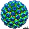
| Virus shell | Shell ID: 1 / Name: VP1 / Diameter: 520 Å / T number (triangulation number): 7 |
-Experimental details
-Structure determination
| Method | cryo EM |
|---|---|
 Processing Processing | single particle reconstruction |
| Aggregation state | particle |
-Sample preparation
| Vitrification | Cryogen name: ETHANE / Instrument: OTHER / Details: Vitrification instrument: Vitrobot |
|---|
- Electron microscopy
| Microscope | FEI TECNAI F30 |
|---|---|
| Image recording | Category: FILM / Film or detector model: KODAK SO-163 FILM / Digitization - Scanner: NIKON SUPER COOLSCAN 9000 / Digitization - Sampling interval: 6.3 µm |
| Electron beam | Acceleration voltage: 200 kV / Electron source:  FIELD EMISSION GUN FIELD EMISSION GUN |
| Electron optics | Illumination mode: FLOOD BEAM / Imaging mode: BRIGHT FIELD / Nominal defocus max: 4.9 µm / Nominal defocus min: 0.7 µm / Nominal magnification: 39000 |
| Sample stage | Specimen holder: Eucentric / Specimen holder model: GATAN LIQUID NITROGEN |
| Experimental equipment |  Model: Tecnai F30 / Image courtesy: FEI Company |
-Image processing
| CTF correction | Details: None |
|---|---|
| Final reconstruction | Algorithm: OTHER / Resolution.type: BY AUTHOR / Resolution: 35.0 Å / Resolution method: FSC 0.5 CUT-OFF / Software - Name: PFT3DR, Bsoft Details: Random-model method. Angular step-size was initially set to 20 deg. in the first iteration and gradually decreased by 0.19 deg. in each successive iteration, until a lower limit of 1 deg. was reached. Number images used: 4198 |
