 Movie
Movie Controller


+ Open data

- Basic information
| Entry | Database: PDB / ID: 1k4r | ||||||
|---|---|---|---|---|---|---|---|
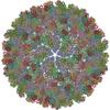
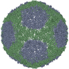
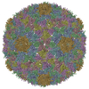
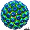
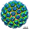
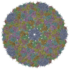
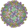
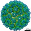
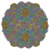
| Title | Structure of Dengue Virus | ||||||
 Components Components | MAJOR ENVELOPE PROTEIN E | ||||||
 Keywords Keywords | VIRUS / Flavivirus / Flaviviridae / Dengue virus / glycoprotein E from tick-borne encephalitis virus / Icosahedral virus | ||||||
| Function / homology |  Function and homology information Function and homology informationsymbiont-mediated suppression of host JAK-STAT cascade via inhibition of STAT2 activity / viral capsid / ribonucleoside triphosphate phosphatase activity / double-stranded RNA binding / methyltransferase cap1 activity / mRNA 5'-cap (guanine-N7-)-methyltransferase activity / RNA helicase activity / protein dimerization activity / symbiont-mediated suppression of host innate immune response / host cell endoplasmic reticulum membrane ...symbiont-mediated suppression of host JAK-STAT cascade via inhibition of STAT2 activity / viral capsid / ribonucleoside triphosphate phosphatase activity / double-stranded RNA binding / methyltransferase cap1 activity / mRNA 5'-cap (guanine-N7-)-methyltransferase activity / RNA helicase activity / protein dimerization activity / symbiont-mediated suppression of host innate immune response / host cell endoplasmic reticulum membrane / symbiont-mediated suppression of host type I interferon-mediated signaling pathway / serine-type endopeptidase activity / viral RNA genome replication / RNA-directed RNA polymerase activity / fusion of virus membrane with host endosome membrane / symbiont entry into host cell / virion attachment to host cell / host cell nucleus / virion membrane / structural molecule activity / proteolysis / extracellular region / ATP binding / membrane / metal ion binding Similarity search - Function | ||||||
| Biological species |  Chimeric Tick-borne encephalitis virus/Dengue virus 4 Chimeric Tick-borne encephalitis virus/Dengue virus 4 | ||||||
| Method | ELECTRON MICROSCOPY / single particle reconstruction / cryo EM / Resolution: 24 Å | ||||||
 Authors Authors | Kuhn, R.J. / Zhang, W. / Rossmann, M.G. / Pletnev, S.V. / Corver, J. / Lenches, E. / Jones, C.T. / Mukhopadhyay, S. / Chipman, P.R. / Strauss, E.G. ...Kuhn, R.J. / Zhang, W. / Rossmann, M.G. / Pletnev, S.V. / Corver, J. / Lenches, E. / Jones, C.T. / Mukhopadhyay, S. / Chipman, P.R. / Strauss, E.G. / Baker, T.S. / Strauss, J.H. | ||||||
 Citation Citation | Journal: Cell / Year: 2002 Title: Structure of dengue virus: implications for flavivirus organization, maturation, and fusion. Authors: Richard J Kuhn / Wei Zhang / Michael G Rossmann / Sergei V Pletnev / Jeroen Corver / Edith Lenches / Christopher T Jones / Suchetana Mukhopadhyay / Paul R Chipman / Ellen G Strauss / Timothy ...Authors: Richard J Kuhn / Wei Zhang / Michael G Rossmann / Sergei V Pletnev / Jeroen Corver / Edith Lenches / Christopher T Jones / Suchetana Mukhopadhyay / Paul R Chipman / Ellen G Strauss / Timothy S Baker / James H Strauss /  Abstract: The first structure of a flavivirus has been determined by using a combination of cryoelectron microscopy and fitting of the known structure of glycoprotein E into the electron density map. The virus ...The first structure of a flavivirus has been determined by using a combination of cryoelectron microscopy and fitting of the known structure of glycoprotein E into the electron density map. The virus core, within a lipid bilayer, has a less-ordered structure than the external, icosahedral scaffold of 90 glycoprotein E dimers. The three E monomers per icosahedral asymmetric unit do not have quasiequivalent symmetric environments. Difference maps indicate the location of the small membrane protein M relative to the overlaying scaffold of E dimers. The structure suggests that flaviviruses, and by analogy also alphaviruses, employ a fusion mechanism in which the distal beta barrels of domain II of the glycoprotein E are inserted into the cellular membrane. #1: Journal: Nature / Year: 1995Title: Virology. When It's Better to Lie Low Authors: Kuhn, R.J. / Rossmann, M.G. #2: Journal: Nature / Year: 1995Title: The Envelope Glycoprotein from Tick-borne Encephalitis Virus at 2 A Resolution Authors: Rey, F.A. / Heinz, F.X. / Mandl, C. / Kunz, C. / Harrison, S.C. | ||||||
| History |
| ||||||
| Remark 2 | RESOLUTION. 24.00 ANGSTROM. |
- Structure visualization
| Movie |
Movie viewer |
|---|---|
| Structure viewer | Molecule: MolmilJmol/JSmol |
- Downloads & links
-Download
| PDBx/mmCIF format | 1k4r.cif.gz | 230.2 KB | Display | PDBx/mmCIF format |
|---|---|---|---|---|
| PDB format | pdb1k4r.ent.gz | 186.5 KB | Display | PDB format |
| PDBx/mmJSON format | 1k4r.json.gz | Tree view | PDBx/mmJSON format | |
| Others |  Other downloads Other downloads |
-Validation report
| Arichive directory | https://data.pdbj.org/pub/pdb/validation_reports/k4/1k4rftp://data.pdbj.org/pub/pdb/validation_reports/k4/1k4r | HTTPS FTP |
|---|
-Related structure data
| Related structure data | |
|---|---|
| Similar structure data |


-Links
 PDBj
PDBj

- Assembly
| Deposited unit | 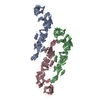
|
|---|---|
| 1 | x 60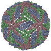
|
| 2 |
|
| 3 | x 5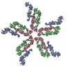
|
| 4 | x 6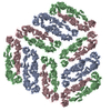
|
| 5 | 
|
| Symmetry | Point symmetry: (Hermann–Mauguin notation: 532 / Schoenflies symbol: I (icosahedral)) |
-Components
| #1: Protein | Mass: 43231.145 Da / Num. of mol.: 3 / Fragment: UNP residues 284-678 / Source method: isolated from a natural source Source: (natural) Chimeric Tick-borne encephalitis virus/Dengue virus 4Genus: Flavivirus / References: UniProt: C3V005 Has protein modification | Y | |
|---|
-Experimental details
-Experiment
| Experiment | Method: ELECTRON MICROSCOPY |
|---|---|
| EM experiment | Aggregation state: PARTICLE / 3D reconstruction method: single particle reconstruction |
- Sample preparation
| Component | Name: DENGUE VIRUS MAJOR ENVELOPE PROTEIN E / Type: VIRUS Details: SAMPLES WERE PREPARED AS THIN LAYERS OF VITREOUS ICE AND MAINTAINED AT NEAR LIQUID NITROGEN TEMPERATURE IN THE ELECTRON MICROSCOPE. |
|---|---|
| Details of virus | Empty: NO / Enveloped: YES / Host category: VERTEBRATES / Isolate: SPECIES / Type: VIRION |
| Natural host | Organism: Primates |
| Buffer solution | pH: 7.6 |
| Specimen | Embedding applied: NO / Shadowing applied: NO / Staining applied: NO / Vitrification applied: YES |
| Specimen support | Details: sample conc. l2.0E10 PFU/ML |
| Vitrification | Details: LIQUID NITROGEN TEMPERATURE 50MM TRIS-HCL, 75MM NACL, 1MM EDTA |
| Crystal grow | *PLUS Method: cryo-electron microscopy |
- Electron microscopy imaging
| Microscopy | Model: FEI/PHILIPS CM200T |
|---|---|
| Electron gun | Electron source:  FIELD EMISSION GUN / Illumination mode: FLOOD BEAM FIELD EMISSION GUN / Illumination mode: FLOOD BEAM |
| Electron lens | Mode: BRIGHT FIELD / Nominal magnification: 50000 X / Nominal defocus max: 1920 nm / Nominal defocus min: 790 nm / Cs: 2 mm |
| Specimen holder | Temperature: 100 K / Tilt angle max: 0 ° / Tilt angle min: 0 ° |
| Image recording | Electron dose: 25.5 e/Å2 / Film or detector model: KODAK SO-163 FILM |
| Image scans | Num. digital images: 25 |
| Reflection | Highest resolution: 24 Å |
- Processing
| Software |
| ||||||||||||
|---|---|---|---|---|---|---|---|---|---|---|---|---|---|
| EM software |
| ||||||||||||
| CTF correction | Details: EACH VIRAL IMAGE WAS CTF CORRECTED BEFORE RECONSTRUCTION, BASED ON THE FOLLOWING EQUATION: F(CORR)=F(OBS)/[|CTF|+WIENER] | ||||||||||||
| Symmetry | Point symmetry: I (icosahedral) | ||||||||||||
| 3D reconstruction | Method: COMMON-LINES AND POLAR-FOURIER-TRANSFORM (FULLER ET AL. 1996, J.STRUC.BIOL. 116, 48-55 BAKER AND CHENG, 1996, J.STRUC.BIOL. 116, 120-130) SOFTWARE USED, PURDUE PROGRAMS Resolution: 24 Å / Num. of particles: 526 / Nominal pixel size: 2.8 Å / Actual pixel size: 2.9 Å / Symmetry type: POINT | ||||||||||||
| Atomic model building | Protocol: RIGID BODY FIT / Space: REAL Details: METHOD--IN THE FIRST STEP THE CRYSTAL STRUCTURE OF TBEV-E CRYSTALLOGRAPHIC DIMER (REY ET.AL., 1995 NATURELONDON) 375, 291-298) WAS ROTATED ABOUT AND TRANSLATEDAROUND AN ICOSAHEDRAL TWO FOLD ...Details: METHOD--IN THE FIRST STEP THE CRYSTAL STRUCTURE OF TBEV-E CRYSTALLOGRAPHIC DIMER (REY ET.AL., 1995 NATURELONDON) 375, 291-298) WAS ROTATED ABOUT AND TRANSLATEDAROUND AN ICOSAHEDRAL TWO FOLD AXES TO FIND THE BEST FIT. AFTER THAT THE DENSITIES AT ALL PIXELS COVERED BY THE FIRST FITTED DIMER WERE SET TO ZERO. THE SECOND DIMER WAS THAN PLACED ON A RADIAL AXIS PASSING THROUGH A POINT NEAR THE QUASI-TWOFOLD AXIS. | ||||||||||||
| Atomic model building | PDB-ID: 1SVB Accession code: 1SVB / Source name: PDB / Type: experimental model | ||||||||||||
| Refinement | Highest resolution: 24 Å Details: Molecules of glycoprotein E of Tick-borne encephalitis virus were solved by using cryo-EM map of Dengue virus | ||||||||||||
| Refinement step | Cycle: LAST / Highest resolution: 24 Å
|
