 Movie
Movie Controller


+ Open data

- Basic information
| Entry | Database: EMDB / ID: EMD-4648 | |||||||||
|---|---|---|---|---|---|---|---|---|---|---|
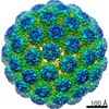
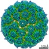
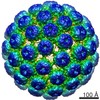
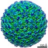
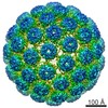
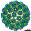
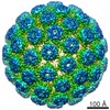
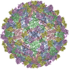
| Title | Packaging of DNA Origami in Viral Capsids | |||||||||
 Map data Map data | Empty capsid from DNA origami-filled SV40 (VP1) preparation. | |||||||||
 Sample Sample |
| |||||||||
| Biological species |  Simian virus 40 Simian virus 40 | |||||||||
| Method | single particle reconstruction / cryo EM / Resolution: 6.8 Å | |||||||||
 Authors Authors | Kopatz I / Zalk R / Levi-Kalisman Y / Zlotkin-Rivkin E / Frank GA / Kler S | |||||||||
 Citation Citation | Journal: Nanoscale / Year: 2019 Title: Packaging of DNA origami in viral capsids. Authors: Idit Kopatz / Ran Zalk / Yael Levi-Kalisman / Efrat Zlotkin-Rivkin / Gabriel A Frank / Stanislav Kler /  Abstract: Here we show the encapsulation of 35 nm diameter, nearly-spherical, DNA origami by self-assembly of SV40-like (simian virus 40) particles. The self-assembly of this new type of nanoparticles is ...Here we show the encapsulation of 35 nm diameter, nearly-spherical, DNA origami by self-assembly of SV40-like (simian virus 40) particles. The self-assembly of this new type of nanoparticles is highly reproducible and efficient. The structure of these particles was determined by cryo-EM. The capsid forms a regular SV40 lattice of T = 7d icosahedral symmetry and the structural features of encapsulated DNA origami are fully visible. These particles are a promising biomaterial for use in various medical applications. | |||||||||
| History |
|
- Structure visualization
| Movie |
 Movie viewer Movie viewer |
|---|---|
| Structure viewer | EM map: SurfViewMolmilJmol/JSmol |
| Supplemental images |

- Downloads & links
-EMDB archive
| Map data | emd_4648.map.gz | 27.8 MB | EMDB map data format | |
|---|---|---|---|---|
| Header (meta data) | emd-4648-v30.xmlemd-4648.xml | 9.3 KB 9.3 KB | Display Display | EMDB header |
| Images | 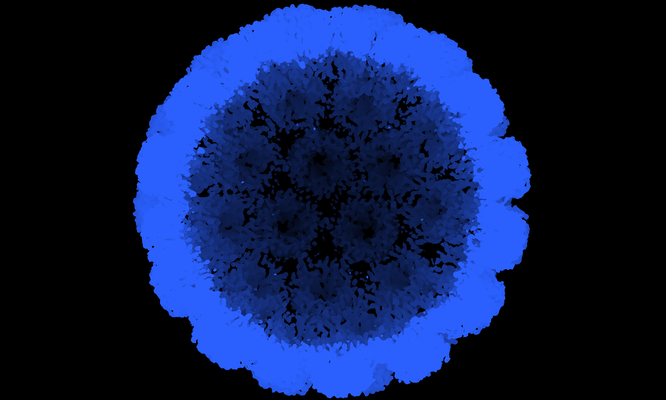 emd_4648.png emd_4648.png | 159.1 KB | ||
| Archive directory |  http://ftp.pdbj.org/pub/emdb/structures/EMD-4648ftp://ftp.pdbj.org/pub/emdb/structures/EMD-4648 http://ftp.pdbj.org/pub/emdb/structures/EMD-4648ftp://ftp.pdbj.org/pub/emdb/structures/EMD-4648 | HTTPS FTP |
-Related structure data


-Links
| EMDB pages | EMDB (EBI/PDBe) / EMDataResource |
|---|---|
| Related items in Molecule of the Month |


-Map
| File | Download / File: emd_4648.map.gz / Format: CCP4 / Size: 103 MB / Type: IMAGE STORED AS FLOATING POINT NUMBER (4 BYTES) | ||||||||||||||||||||||||||||||||||||||||||||||||||||||||||||
|---|---|---|---|---|---|---|---|---|---|---|---|---|---|---|---|---|---|---|---|---|---|---|---|---|---|---|---|---|---|---|---|---|---|---|---|---|---|---|---|---|---|---|---|---|---|---|---|---|---|---|---|---|---|---|---|---|---|---|---|---|---|
| Annotation | Empty capsid from DNA origami-filled SV40 (VP1) preparation. | ||||||||||||||||||||||||||||||||||||||||||||||||||||||||||||
| Projections & slices | Image control
Images are generated by Spider. | ||||||||||||||||||||||||||||||||||||||||||||||||||||||||||||
| Voxel size | X=Y=Z: 2.3 Å | ||||||||||||||||||||||||||||||||||||||||||||||||||||||||||||
| Density |
| ||||||||||||||||||||||||||||||||||||||||||||||||||||||||||||
| Symmetry | Space group: 1 | ||||||||||||||||||||||||||||||||||||||||||||||||||||||||||||
| Details | EMDB XML:
CCP4 map header:
| ||||||||||||||||||||||||||||||||||||||||||||||||||||||||||||
 Z (Sec.)
 Y (Row.)
 X (Col.)


-Supplemental data
- Sample components
-Entire : Simian virus 40
| Entire | Name: Simian virus 40 |
|---|---|
| Components |
|
-Supramolecule #1: Simian virus 40
| Supramolecule | Name: Simian virus 40 / type: virus / ID: 1 / Parent: 0 / NCBI-ID: 10633 / Sci species name: Simian virus 40 / Virus type: VIRUS-LIKE PARTICLE / Virus isolate: OTHER / Virus enveloped: No / Virus empty: No |
|---|---|
| Host system | Organism:   Spodoptera frugiperda (fall armyworm) Spodoptera frugiperda (fall armyworm) |
-Experimental details
-Structure determination
| Method | cryo EM |
|---|---|
 Processing Processing | single particle reconstruction |
| Aggregation state | particle |
-Sample preparation
| Buffer | pH: 7 |
|---|---|
| Grid | Model: Quantifoil R2/2 / Material: COPPER / Mesh: 300 / Support film - Material: CARBON / Support film - topology: HOLEY / Pretreatment - Type: GLOW DISCHARGE |
| Vitrification | Cryogen name: ETHANE / Chamber humidity: 96 % / Chamber temperature: 293 K / Instrument: LEICA EM GP |
- Electron microscopy
| Microscope | FEI POLARA 300 |
|---|---|
| Specialist optics | Energy filter - Name: GIF Quantum LS / Energy filter - Slit width: 20 eV |
| Image recording | Film or detector model: GATAN K2 SUMMIT (4k x 4k) / Detector mode: COUNTING / Average electron dose: 100.0 e/Å2 |
| Electron beam | Acceleration voltage: 300 kV / Electron source:  FIELD EMISSION GUN FIELD EMISSION GUN |
| Electron optics | Illumination mode: FLOOD BEAM / Imaging mode: BRIGHT FIELD |
| Sample stage | Cooling holder cryogen: NITROGEN |
| Experimental equipment |  Model: Tecnai Polara / Image courtesy: FEI Company |
-Image processing
| Final reconstruction | Applied symmetry - Point group: I (icosahedral) / Resolution.type: BY AUTHOR / Resolution: 6.8 Å / Resolution method: FSC 0.143 CUT-OFF / Software - Name: RELION (ver. 3.0 beta2) / Number images used: 13818 |
|---|---|
| Initial angle assignment | Type: MAXIMUM LIKELIHOOD |
| Final angle assignment | Type: MAXIMUM LIKELIHOOD |
-Atomic model buiding 1
| Refinement | Protocol: AB INITIO MODEL |
|---|
