Movie
Movie Controller

[English] 日本語
 Yorodumi
- EMDB-4400: CryoEM reconstruction of full-length, fully-glycosylated human bu... -
+ Open data

- Basic information
| Entry | Database: EMDB / ID: EMD-4400 | |||||||||
|---|---|---|---|---|---|---|---|---|---|---|
| Title | CryoEM reconstruction of full-length, fully-glycosylated human butyrylcholinesterase tetramer | |||||||||
 Map data Map data | cryo-EM map of tetrameric full-length, glycosylated human butyrylcholinesterase (HuBChE) purified from human plasma | |||||||||
 Sample Sample |
| |||||||||
 Keywords Keywords | cholinesterase / tetramer / HYDROLASE | |||||||||
| Function / homology |  Function and homology information Function and homology informationcholinesterase / neuroblast differentiation / cocaine catabolic process / response to folic acid / Neurotransmitter clearance / choline binding / acetylcholine catabolic process / response to alkaloid / cholinesterase activity / choline metabolic process ...cholinesterase / neuroblast differentiation / cocaine catabolic process / response to folic acid / Neurotransmitter clearance / choline binding / acetylcholine catabolic process / response to alkaloid / cholinesterase activity / choline metabolic process / negative regulation of synaptic transmission / peptide hormone processing / hydrolase activity, acting on ester bonds / acetylcholinesterase activity / Aspirin ADME / nuclear envelope lumen / Synthesis of PC / catalytic activity / Synthesis, secretion, and deacylation of Ghrelin / xenobiotic metabolic process / response to glucocorticoid / learning / amyloid-beta binding / blood microparticle / endoplasmic reticulum lumen / negative regulation of cell population proliferation / enzyme binding / : / extracellular region / identical protein binding / plasma membrane Similarity search - Function | |||||||||
| Biological species |  Homo sapiens (human) Homo sapiens (human) | |||||||||
| Method | single particle reconstruction / cryo EM / Resolution: 5.7 Å | |||||||||
 Authors Authors | Leung MR / van Bezouwen LS | |||||||||
 Citation Citation | Journal: Proc Natl Acad Sci U S A / Year: 2018 Title: Cryo-EM structure of the native butyrylcholinesterase tetramer reveals a dimer of dimers stabilized by a superhelical assembly. Authors: Miguel Ricardo Leung / Laura S van Bezouwen / Lawrence M Schopfer / Joel L Sussman / Israel Silman / Oksana Lockridge / Tzviya Zeev-Ben-Mordehai /     Abstract: The quaternary structures of the cholinesterases, acetylcholinesterase (AChE) and butyrylcholinesterase (BChE), are essential for their localization and function. Of practical importance, BChE is a ...The quaternary structures of the cholinesterases, acetylcholinesterase (AChE) and butyrylcholinesterase (BChE), are essential for their localization and function. Of practical importance, BChE is a promising therapeutic candidate for intoxication by organophosphate nerve agents and insecticides, and for detoxification of addictive substances. Efficacy of the recombinant enzyme hinges on its having a long circulatory half-life; this, in turn, depends strongly on its ability to tetramerize. Here, we used cryoelectron microscopy (cryo-EM) to determine the structure of the highly glycosylated native BChE tetramer purified from human plasma at 5.7 Å. Our structure reveals that the BChE tetramer is organized as a staggered dimer of dimers. Tetramerization is mediated by assembly of the C-terminal tryptophan amphiphilic tetramerization (WAT) helices from each subunit as a superhelical assembly around a central lamellipodin-derived oligopeptide with a proline-rich attachment domain (PRAD) sequence that adopts a polyproline II helical conformation and runs antiparallel. The catalytic domains within a dimer are asymmetrically linked to the WAT/PRAD. In the resulting arrangement, the tetramerization domain is largely shielded by the catalytic domains, which may contribute to the stability of the human BChE (HuBChE) tetramer. Our cryo-EM structure reveals the basis for assembly of the native tetramers and has implications for the therapeutic applications of HuBChE. This mode of tetramerization is seen only in the cholinesterases but may provide a promising template for designing other proteins with improved circulatory residence times. | |||||||||
| History |
|
- Structure visualization
| Movie |
Movie viewer |
|---|---|
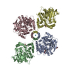
| Structure viewer | EM map: SurfViewMolmilJmol/JSmol |
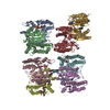
| Supplemental images |

- Downloads & links
-EMDB archive
| Map data | emd_4400.map.gz | 8.4 MB | EMDB map data format | |
|---|---|---|---|---|
| Header (meta data) | emd-4400-v30.xmlemd-4400.xml | 14.7 KB 14.7 KB | Display Display | EMDB header |
| FSC (resolution estimation) | emd_4400_fsc.xml | 10.8 KB | Display | FSC data file |
| Images | 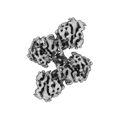 emd_4400.png emd_4400.png | 37.6 KB | ||
| Filedesc metadata | emd-4400.cif.gz | 6.2 KB | ||
| Archive directory |  http://ftp.pdbj.org/pub/emdb/structures/EMD-4400ftp://ftp.pdbj.org/pub/emdb/structures/EMD-4400 http://ftp.pdbj.org/pub/emdb/structures/EMD-4400ftp://ftp.pdbj.org/pub/emdb/structures/EMD-4400 | HTTPS FTP |
-Related structure data
| Related structure data |  6i2tMC M: atomic model generated by this map C: citing same article ( |
|---|---|
| Similar structure data |


-Links
| EMDB pages | EMDB (EBI/PDBe) / EMDataResource |
|---|---|
| Related items in Molecule of the Month |


-Map
| File | Download / File: emd_4400.map.gz / Format: CCP4 / Size: 103 MB / Type: IMAGE STORED AS FLOATING POINT NUMBER (4 BYTES) | ||||||||||||||||||||||||||||||||||||||||||||||||||||||||||||
|---|---|---|---|---|---|---|---|---|---|---|---|---|---|---|---|---|---|---|---|---|---|---|---|---|---|---|---|---|---|---|---|---|---|---|---|---|---|---|---|---|---|---|---|---|---|---|---|---|---|---|---|---|---|---|---|---|---|---|---|---|---|
| Annotation | cryo-EM map of tetrameric full-length, glycosylated human butyrylcholinesterase (HuBChE) purified from human plasma | ||||||||||||||||||||||||||||||||||||||||||||||||||||||||||||
| Projections & slices | Image control
Images are generated by Spider. | ||||||||||||||||||||||||||||||||||||||||||||||||||||||||||||
| Voxel size | X=Y=Z: 1.0285 Å | ||||||||||||||||||||||||||||||||||||||||||||||||||||||||||||
| Density |
| ||||||||||||||||||||||||||||||||||||||||||||||||||||||||||||
| Symmetry | Space group: 1 | ||||||||||||||||||||||||||||||||||||||||||||||||||||||||||||
| Details | EMDB XML:
CCP4 map header:
| ||||||||||||||||||||||||||||||||||||||||||||||||||||||||||||
 Z (Sec.)
 Y (Row.)
 X (Col.)


-Supplemental data
- Sample components
-Entire : heteropentameric complex consisting of four copies of butyrylchol...
| Entire | Name: heteropentameric complex consisting of four copies of butyrylcholinesterase and one copy of a lamellipodin-derived polyproline peptide |
|---|---|
| Components |
|
-Supramolecule #1: heteropentameric complex consisting of four copies of butyrylchol...
| Supramolecule | Name: heteropentameric complex consisting of four copies of butyrylcholinesterase and one copy of a lamellipodin-derived polyproline peptide type: complex / ID: 1 / Parent: 0 / Macromolecule list: #1-#2 |
|---|---|
| Source (natural) | Organism: Homo sapiens (human) / Tissue: plasma |
| Molecular weight | Theoretical: 340 KDa |
-Macromolecule #1: Cholinesterase
| Macromolecule | Name: Cholinesterase / type: protein_or_peptide / ID: 1 / Number of copies: 4 / Enantiomer: LEVO / EC number: cholinesterase |
|---|---|
| Source (natural) | Organism: Homo sapiens (human) |
| Molecular weight | Theoretical: 65.1495 KDa |
| Sequence | String: EDDIIIATKN GKVRGMNLTV FGGTVTAFLG IPYAQPPLGR LRFKKPQSLT KWSDIWNATK YANSCCQNID QSFPGFHGSE MWNPNTDLS EDCLYLNVWI PAPKPKNATV LIWIYGGGFQ TGTSSLHVYD GKFLARVERV IVVSMNYRVG ALGFLALPGN P EAPGNMGL ...String: EDDIIIATKN GKVRGMNLTV FGGTVTAFLG IPYAQPPLGR LRFKKPQSLT KWSDIWNATK YANSCCQNID QSFPGFHGSE MWNPNTDLS EDCLYLNVWI PAPKPKNATV LIWIYGGGFQ TGTSSLHVYD GKFLARVERV IVVSMNYRVG ALGFLALPGN P EAPGNMGL FDQQLALQWV QKNIAAFGGN PKSVTLFGES AGAASVSLHL LSPGSHSLFT RAILQSGSFN APWAVTSLYE AR NRTLNLA KLTGCSRENE TEIIKCLRNK DPQEILLNEA FVVPYGTPLS VNFGPTVDGD FLTDMPDILL ELGQFKKTQI LVG VNKDEG TAFLVYGAPG FSKDNNSIIT RKEFQEGLKI FFPGVSEFGK ESILFHYTDW VDDQRPENYR EALGDVVGDY NFIC PALEF TKKFSEWGNN AFFYYFEHRS SKLPWPEWMG VMHGYEIEFV FGLPLERRDN YTKAEEILSR SIVKRWANFA KYGNP NETQ NNSTSWPVFK STEQKYLTLN TESTRIMTKL RAQQCRFWTS FFPKVLEMTG NIDEAEWEWK AGFHRWNNYM MDWKNQ FND YTSKKESCVG L UniProtKB: Cholinesterase |
-Macromolecule #2: lamellipodin-derived polyproline peptide
| Macromolecule | Name: lamellipodin-derived polyproline peptide / type: protein_or_peptide / ID: 2 / Number of copies: 1 / Enantiomer: LEVO |
|---|---|
| Source (natural) | Organism: Homo sapiens (human) |
| Molecular weight | Theoretical: 1.183393 KDa |
| Sequence | String: PPPPPPPPPP PP |
-Macromolecule #3: 2-acetamido-2-deoxy-beta-D-glucopyranose
| Macromolecule | Name: 2-acetamido-2-deoxy-beta-D-glucopyranose / type: ligand / ID: 3 / Number of copies: 8 / Formula: NAG |
|---|---|
| Molecular weight | Theoretical: 221.208 Da |
| Chemical component information |  ChemComp-NAG: |
-Experimental details
-Structure determination
| Method | cryo EM |
|---|---|
 Processing Processing | single particle reconstruction |
| Aggregation state | particle |
-Sample preparation
| Concentration | 3.3 mg/mL |
|---|---|
| Buffer | pH: 8 / Component - Concentration: 10.0 mM / Component - Formula: C4H11NO3 / Component - Name: Tris |
| Grid | Model: Quantifoil R1.2/1.3 / Material: COPPER / Mesh: 300 / Support film - Material: CARBON / Support film - topology: HOLEY / Pretreatment - Type: GLOW DISCHARGE / Pretreatment - Time: 30 sec. |
| Vitrification | Cryogen name: ETHANE / Chamber humidity: 90 % / Chamber temperature: 293 K / Instrument: FEI VITROBOT MARK IV Details: blot for either 2s or 3s at blot force -2 or 0, respectively. |
- Electron microscopy
| Microscope | FEI TALOS ARCTICA |
|---|---|
| Specialist optics | Energy filter - Name: GIF Quantum LS / Energy filter - Slit width: 20 eV |
| Image recording | Film or detector model: GATAN K2 SUMMIT (4k x 4k) / Detector mode: SUPER-RESOLUTION / Number grids imaged: 2 / Number real images: 4518 / Average exposure time: 6.3 sec. / Average electron dose: 49.5 e/Å2 |
| Electron beam | Acceleration voltage: 200 kV / Electron source:  FIELD EMISSION GUN FIELD EMISSION GUN |
| Electron optics | C2 aperture diameter: 50.0 µm / Illumination mode: FLOOD BEAM / Imaging mode: BRIGHT FIELD / Cs: 2.7 mm / Nominal defocus max: 2.5 µm / Nominal defocus min: 1.2 µm / Nominal magnification: 130000 |
| Sample stage | Specimen holder model: OTHER / Cooling holder cryogen: NITROGEN |
| Experimental equipment |  Model: Talos Arctica / Image courtesy: FEI Company |
+Image processing

-Atomic model buiding 1
| Initial model |
| ||||||
|---|---|---|---|---|---|---|---|
| Refinement | Space: REAL / Protocol: RIGID BODY FIT | ||||||
| Output model | PDB-6i2t: |