 Movie
Movie Controller

[English] 日本語
 Yorodumi
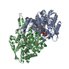
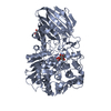
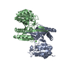
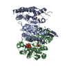
- PDB-6kdy: Crystal structure of the alpha bata heterodimer of human IDH3 in ... -
+ Open data

- Basic information
| Entry | Database: PDB / ID: 6kdy | ||||||
|---|---|---|---|---|---|---|---|
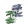
| Title | Crystal structure of the alpha bata heterodimer of human IDH3 in complex with NAD. | ||||||
 Components Components | (Isocitrate dehydrogenase [NAD] subunit ...) x 2 | ||||||
 Keywords Keywords | OXIDOREDUCTASE / NAD dependent isocitrate dehydrogenase | ||||||
| Function / homology |  Function and homology information Function and homology informationisocitrate dehydrogenase complex (NAD+) / isocitrate dehydrogenase (NAD+) / isocitrate dehydrogenase (NAD+) activity / Citric acid cycle (TCA cycle) / isocitrate metabolic process / tricarboxylic acid cycle / Mitochondrial protein degradation / NAD binding / carbohydrate metabolic process / electron transfer activity ...isocitrate dehydrogenase complex (NAD+) / isocitrate dehydrogenase (NAD+) / isocitrate dehydrogenase (NAD+) activity / Citric acid cycle (TCA cycle) / isocitrate metabolic process / tricarboxylic acid cycle / Mitochondrial protein degradation / NAD binding / carbohydrate metabolic process / electron transfer activity / mitochondrial matrix / magnesium ion binding / mitochondrion / nucleus Similarity search - Function | ||||||
| Biological species |  Homo sapiens (human) Homo sapiens (human) | ||||||
| Method |  X-RAY DIFFRACTION / SYNCHROTRON / MOLECULAR REPLACEMENT / Resolution: 3.02 Å X-RAY DIFFRACTION / SYNCHROTRON / MOLECULAR REPLACEMENT / Resolution: 3.02 Å | ||||||
 Authors Authors | Sun, P. / Ding, J. | ||||||
| Funding support |  China, 1items China, 1items
| ||||||
 Citation Citation | Journal: J.Biol.Chem. / Year: 2019 Title: Molecular basis for the function of the alpha beta heterodimer of human NAD-dependent isocitrate dehydrogenase. Authors: Sun, P. / Ma, T. / Zhang, T. / Zhu, H. / Zhang, J. / Liu, Y. / Ding, J. | ||||||
| History |
|
- Structure visualization
| Structure viewer | Molecule: MolmilJmol/JSmol |
|---|
- Downloads & links
-Download
| PDBx/mmCIF format | 6kdy.cif.gz | 505.9 KB | Display | PDBx/mmCIF format |
|---|---|---|---|---|
| PDB format | pdb6kdy.ent.gz | 409.7 KB | Display | PDB format |
| PDBx/mmJSON format | 6kdy.json.gz | Tree view | PDBx/mmJSON format | |
| Others |  Other downloads Other downloads |
-Validation report
| Arichive directory | https://data.pdbj.org/pub/pdb/validation_reports/kd/6kdyftp://data.pdbj.org/pub/pdb/validation_reports/kd/6kdy | HTTPS FTP |
|---|
-Related structure data
| Related structure data |  6kdeSC  6kdfC  6ke3C S: Starting model for refinement C: citing same article ( |
|---|---|
| Similar structure data |


-Links
 PDBj
PDBj

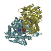
- Assembly
| Deposited unit | 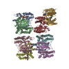
| ||||||||
|---|---|---|---|---|---|---|---|---|---|
| 1 | 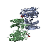
| ||||||||
| 2 | 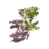
| ||||||||
| 3 | 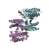
| ||||||||
| 4 | 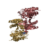
| ||||||||
| Unit cell |
|
-Components
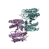
-Isocitrate dehydrogenase [NAD] subunit ... , 2 types, 8 molecules ACEGBDFH
| #1: Protein | Mass: 36826.305 Da / Num. of mol.: 4 Source method: isolated from a genetically manipulated source Source: (gene. exp.) Homo sapiens (human) / Gene: IDH3A / Production host:  References: UniProt: P50213, isocitrate dehydrogenase (NAD+) #2: Protein | Mass: 39703.664 Da / Num. of mol.: 4 Source method: isolated from a genetically manipulated source Source: (gene. exp.) Homo sapiens (human) / Gene: IDH3B / Production host: |
|---|
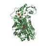
-Non-polymers , 5 types, 9 molecules 


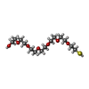

| #3: Chemical | ChemComp-NAD /  Mass: 663.425 Da / Num. of mol.: 4 / Source method: obtained synthetically / Formula: C21H27N7O14P2 / Feature type: SUBJECT OF INVESTIGATION / Comment: NAD*YM Mass: 663.425 Da / Num. of mol.: 4 / Source method: obtained synthetically / Formula: C21H27N7O14P2 / Feature type: SUBJECT OF INVESTIGATION / Comment: NAD*YM#4: Chemical |  Mass: 194.226 Da / Num. of mol.: 2 / Source method: obtained synthetically / Formula: C8H18O5 / Comment: precipitant*YM Mass: 194.226 Da / Num. of mol.: 2 / Source method: obtained synthetically / Formula: C8H18O5 / Comment: precipitant*YM#5: Chemical | ChemComp-2PE / |  Mass: 414.488 Da / Num. of mol.: 1 / Source method: obtained synthetically / Formula: C18H38O10 / Comment: precipitant*YM Mass: 414.488 Da / Num. of mol.: 1 / Source method: obtained synthetically / Formula: C18H38O10 / Comment: precipitant*YM#6: Chemical | ChemComp-PE7 / |  Mass: 342.449 Da / Num. of mol.: 1 / Source method: obtained synthetically / Formula: C14H30O7S Mass: 342.449 Da / Num. of mol.: 1 / Source method: obtained synthetically / Formula: C14H30O7S#7: Chemical | ChemComp-P6G / |  Mass: 282.331 Da / Num. of mol.: 1 / Source method: obtained synthetically / Formula: C12H26O7 / Comment: precipitant*YM Mass: 282.331 Da / Num. of mol.: 1 / Source method: obtained synthetically / Formula: C12H26O7 / Comment: precipitant*YM |
|---|
-Details
| Has ligand of interest | Y |
|---|---|
| Sequence details | THIS SEQUENCE OF IDH3 B CORRESPOND |
-Experimental details
-Experiment
| Experiment | Method: X-RAY DIFFRACTION / Number of used crystals: 1 |
|---|
- Sample preparation
| Crystal | Density Matthews: 3.07 Å3/Da / Density % sol: 60 % |
|---|---|
| Crystal grow | Temperature: 289 K / Method: vapor diffusion, hanging drop Details: 0.05M ICT, 0.05M NAD, 0.2 M calcium acetate (pH 7.5) and 20% (w/v) PEG3350 |
-Data collection
| Diffraction | Mean temperature: 100 K / Serial crystal experiment: N | |||||||||||||||||||||||||||||||||||||||||||||||||||||||||||||||||||||||||||||||||||||||||||||||||||
|---|---|---|---|---|---|---|---|---|---|---|---|---|---|---|---|---|---|---|---|---|---|---|---|---|---|---|---|---|---|---|---|---|---|---|---|---|---|---|---|---|---|---|---|---|---|---|---|---|---|---|---|---|---|---|---|---|---|---|---|---|---|---|---|---|---|---|---|---|---|---|---|---|---|---|---|---|---|---|---|---|---|---|---|---|---|---|---|---|---|---|---|---|---|---|---|---|---|---|---|---|
| Diffraction source | Source: SYNCHROTRON / Site: SSRF / Beamline: BL17U1 / Wavelength: 0.9792 Å | |||||||||||||||||||||||||||||||||||||||||||||||||||||||||||||||||||||||||||||||||||||||||||||||||||
| Detector | Type: DECTRIS PILATUS3 6M / Detector: PIXEL / Date: Feb 1, 2019 | |||||||||||||||||||||||||||||||||||||||||||||||||||||||||||||||||||||||||||||||||||||||||||||||||||
| Radiation | Protocol: SINGLE WAVELENGTH / Monochromatic (M) / Laue (L): M / Scattering type: x-ray | |||||||||||||||||||||||||||||||||||||||||||||||||||||||||||||||||||||||||||||||||||||||||||||||||||
| Radiation wavelength | Wavelength: 0.9792 Å / Relative weight: 1 | |||||||||||||||||||||||||||||||||||||||||||||||||||||||||||||||||||||||||||||||||||||||||||||||||||
| Reflection | Resolution: 3.001→50 Å / Num. obs: 71108 / % possible obs: 99.8 % / Redundancy: 3.4 % / Rmerge(I) obs: 0.08 / Rpim(I) all: 0.051 / Rrim(I) all: 0.095 / Χ2: 1.062 / Net I/σ(I): 8.5 | |||||||||||||||||||||||||||||||||||||||||||||||||||||||||||||||||||||||||||||||||||||||||||||||||||
| Reflection shell | Diffraction-ID: 1
|
- Processing
| Software |
| ||||||||||||||||||||||||||||||||||||||||||||||||||||||||||||||||||||||||||||||||||||||||||||||||||||||||||||||||||||||||||||||||||||||||||||||||||||||||||||||||||
|---|---|---|---|---|---|---|---|---|---|---|---|---|---|---|---|---|---|---|---|---|---|---|---|---|---|---|---|---|---|---|---|---|---|---|---|---|---|---|---|---|---|---|---|---|---|---|---|---|---|---|---|---|---|---|---|---|---|---|---|---|---|---|---|---|---|---|---|---|---|---|---|---|---|---|---|---|---|---|---|---|---|---|---|---|---|---|---|---|---|---|---|---|---|---|---|---|---|---|---|---|---|---|---|---|---|---|---|---|---|---|---|---|---|---|---|---|---|---|---|---|---|---|---|---|---|---|---|---|---|---|---|---|---|---|---|---|---|---|---|---|---|---|---|---|---|---|---|---|---|---|---|---|---|---|---|---|---|---|---|---|---|---|---|
| Refinement | Method to determine structure: MOLECULAR REPLACEMENT Starting model: 6KDE Resolution: 3.02→48.928 Å / SU ML: 0.41 / Cross valid method: THROUGHOUT / σ(F): 1.33 / Phase error: 26.44
| ||||||||||||||||||||||||||||||||||||||||||||||||||||||||||||||||||||||||||||||||||||||||||||||||||||||||||||||||||||||||||||||||||||||||||||||||||||||||||||||||||
| Solvent computation | Shrinkage radii: 0.9 Å / VDW probe radii: 1.11 Å | ||||||||||||||||||||||||||||||||||||||||||||||||||||||||||||||||||||||||||||||||||||||||||||||||||||||||||||||||||||||||||||||||||||||||||||||||||||||||||||||||||
| Displacement parameters | Biso max: 243.45 Å2 / Biso mean: 71.4049 Å2 / Biso min: 26.83 Å2 | ||||||||||||||||||||||||||||||||||||||||||||||||||||||||||||||||||||||||||||||||||||||||||||||||||||||||||||||||||||||||||||||||||||||||||||||||||||||||||||||||||
| Refinement step | Cycle: final / Resolution: 3.02→48.928 Å
| ||||||||||||||||||||||||||||||||||||||||||||||||||||||||||||||||||||||||||||||||||||||||||||||||||||||||||||||||||||||||||||||||||||||||||||||||||||||||||||||||||
| LS refinement shell | Refine-ID: X-RAY DIFFRACTION / Rfactor Rfree error: 0
|
