Movie
Movie Controller

[English] 日本語
 Yorodumi
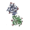
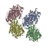
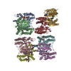
- PDB-6i2t: CryoEM reconstruction of full-length, fully-glycosylated human bu... -
+ Open data

- Basic information
| Entry | Database: PDB / ID: 6i2t | ||||||
|---|---|---|---|---|---|---|---|
| Title | CryoEM reconstruction of full-length, fully-glycosylated human butyrylcholinesterase tetramer | ||||||
 Components Components |
| ||||||
 Keywords Keywords | HYDROLASE / cholinesterase / tetramer | ||||||
| Function / homology |  Function and homology information Function and homology informationcholinesterase / neuroblast differentiation / cocaine catabolic process / response to folic acid / Neurotransmitter clearance / choline binding / acetylcholine catabolic process / response to alkaloid / cholinesterase activity / choline metabolic process ...cholinesterase / neuroblast differentiation / cocaine catabolic process / response to folic acid / Neurotransmitter clearance / choline binding / acetylcholine catabolic process / response to alkaloid / cholinesterase activity / choline metabolic process / negative regulation of synaptic transmission / peptide hormone processing / hydrolase activity, acting on ester bonds / acetylcholinesterase activity / Aspirin ADME / nuclear envelope lumen / Synthesis of PC / catalytic activity / Synthesis, secretion, and deacylation of Ghrelin / xenobiotic metabolic process / response to glucocorticoid / learning / amyloid-beta binding / blood microparticle / endoplasmic reticulum lumen / negative regulation of cell population proliferation / enzyme binding / : / extracellular region / identical protein binding / plasma membrane Similarity search - Function | ||||||
| Biological species |  Homo sapiens (human) Homo sapiens (human) | ||||||
| Method | ELECTRON MICROSCOPY / single particle reconstruction / cryo EM / Resolution: 5.7 Å | ||||||
 Authors Authors | Leung, M.R. / van Bezouwen, L.S. / Schopfer, L.M. / Sussman, J.L. / Silman, I. / Lockridge, O. / Zeev-Ben-Mordehai, T. | ||||||
 Citation Citation | Journal: Proc Natl Acad Sci U S A / Year: 2018 Title: Cryo-EM structure of the native butyrylcholinesterase tetramer reveals a dimer of dimers stabilized by a superhelical assembly. Authors: Miguel Ricardo Leung / Laura S van Bezouwen / Lawrence M Schopfer / Joel L Sussman / Israel Silman / Oksana Lockridge / Tzviya Zeev-Ben-Mordehai /     Abstract: The quaternary structures of the cholinesterases, acetylcholinesterase (AChE) and butyrylcholinesterase (BChE), are essential for their localization and function. Of practical importance, BChE is a ...The quaternary structures of the cholinesterases, acetylcholinesterase (AChE) and butyrylcholinesterase (BChE), are essential for their localization and function. Of practical importance, BChE is a promising therapeutic candidate for intoxication by organophosphate nerve agents and insecticides, and for detoxification of addictive substances. Efficacy of the recombinant enzyme hinges on its having a long circulatory half-life; this, in turn, depends strongly on its ability to tetramerize. Here, we used cryoelectron microscopy (cryo-EM) to determine the structure of the highly glycosylated native BChE tetramer purified from human plasma at 5.7 Å. Our structure reveals that the BChE tetramer is organized as a staggered dimer of dimers. Tetramerization is mediated by assembly of the C-terminal tryptophan amphiphilic tetramerization (WAT) helices from each subunit as a superhelical assembly around a central lamellipodin-derived oligopeptide with a proline-rich attachment domain (PRAD) sequence that adopts a polyproline II helical conformation and runs antiparallel. The catalytic domains within a dimer are asymmetrically linked to the WAT/PRAD. In the resulting arrangement, the tetramerization domain is largely shielded by the catalytic domains, which may contribute to the stability of the human BChE (HuBChE) tetramer. Our cryo-EM structure reveals the basis for assembly of the native tetramers and has implications for the therapeutic applications of HuBChE. This mode of tetramerization is seen only in the cholinesterases but may provide a promising template for designing other proteins with improved circulatory residence times. | ||||||
| History |
|
- Structure visualization
| Movie |
Movie viewer |
|---|---|
| Structure viewer | Molecule: MolmilJmol/JSmol |
- Downloads & links
-Download
| PDBx/mmCIF format | 6i2t.cif.gz | 394.6 KB | Display | PDBx/mmCIF format |
|---|---|---|---|---|
| PDB format | pdb6i2t.ent.gz | 328.4 KB | Display | PDB format |
| PDBx/mmJSON format | 6i2t.json.gz | Tree view | PDBx/mmJSON format | |
| Others |  Other downloads Other downloads |
-Validation report
| Arichive directory | https://data.pdbj.org/pub/pdb/validation_reports/i2/6i2tftp://data.pdbj.org/pub/pdb/validation_reports/i2/6i2t | HTTPS FTP |
|---|
-Related structure data
| Related structure data |  4400MC M: map data used to model this data C: citing same article ( |
|---|---|
| Similar structure data |


-Links
 PDBj
PDBj


- Assembly
| Deposited unit | 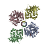
|
|---|---|
| 1 |
|
-Components
| #1: Protein | Mass: 65149.500 Da / Num. of mol.: 4 / Source method: isolated from a natural source / Source: (natural) Homo sapiens (human) / References: UniProt: P06276, cholinesterase#2: Protein/peptide | | Mass: 1183.393 Da / Num. of mol.: 1 / Source method: isolated from a natural source / Source: (natural) Homo sapiens (human)#3: Sugar | ChemComp-NAG /   Type: D-saccharide, beta linking / Mass: 221.208 Da / Num. of mol.: 8 Type: D-saccharide, beta linking / Mass: 221.208 Da / Num. of mol.: 8Source method: isolated from a genetically manipulated source Formula: C8H15NO6 Has protein modification | Y | |
|---|
-Experimental details
-Experiment
| Experiment | Method: ELECTRON MICROSCOPY |
|---|---|
| EM experiment | Aggregation state: PARTICLE / 3D reconstruction method: single particle reconstruction |
- Sample preparation
| Component | Name: heteropentameric complex consisting of four copies of butyrylcholinesterase and one copy of a lamellipodin-derived polyproline peptide Type: COMPLEX / Entity ID: #1-#2 / Source: NATURAL |
|---|---|
| Molecular weight | Value: 0.34 MDa / Experimental value: YES |
| Source (natural) | Organism: Homo sapiens (human) / Tissue: plasma |
| Buffer solution | pH: 8 |
| Buffer component | Conc.: 10 mM / Name: Tris / Formula: C4H11NO3 |
| Specimen | Conc.: 3.3 mg/ml / Embedding applied: NO / Shadowing applied: NO / Staining applied: NO / Vitrification applied: YES |
| Specimen support | Grid material: COPPER / Grid mesh size: 300 divisions/in. / Grid type: Quantifoil R1.2/1.3 |
| Vitrification | Instrument: FEI VITROBOT MARK IV / Cryogen name: ETHANE / Humidity: 90 % / Chamber temperature: 293 K Details: blot for either 2s or 3s at blot force -2 or 0, respectively |
- Electron microscopy imaging
| Experimental equipment |  Model: Talos Arctica / Image courtesy: FEI Company |
|---|---|
| Microscopy | Model: FEI TALOS ARCTICA |
| Electron gun | Electron source:  FIELD EMISSION GUN / Accelerating voltage: 200 kV / Illumination mode: FLOOD BEAM FIELD EMISSION GUN / Accelerating voltage: 200 kV / Illumination mode: FLOOD BEAM |
| Electron lens | Mode: BRIGHT FIELD / Nominal magnification: 130000 X / Nominal defocus max: 2500 nm / Nominal defocus min: 1200 nm / Cs: 2.7 mm / C2 aperture diameter: 50 µm |
| Specimen holder | Cryogen: NITROGEN / Specimen holder model: OTHER |
| Image recording | Average exposure time: 6.3 sec. / Electron dose: 49.5 e/Å2 / Detector mode: SUPER-RESOLUTION / Film or detector model: GATAN K2 SUMMIT (4k x 4k) / Num. of grids imaged: 2 / Num. of real images: 4518 |
| EM imaging optics | Energyfilter name: GIF Quantum LS / Energyfilter slit width: 20 eV |
- Processing
| Software | Name: PHENIX / Version: 1.14_3260: / Classification: refinement | |||||||||||||||||||||||||||
|---|---|---|---|---|---|---|---|---|---|---|---|---|---|---|---|---|---|---|---|---|---|---|---|---|---|---|---|---|
| EM software |
| |||||||||||||||||||||||||||
| CTF correction | Type: PHASE FLIPPING AND AMPLITUDE CORRECTION | |||||||||||||||||||||||||||
| Particle selection | Num. of particles selected: 414189 | |||||||||||||||||||||||||||
| Symmetry | Point symmetry: C2 (2 fold cyclic) | |||||||||||||||||||||||||||
| 3D reconstruction | Resolution: 5.7 Å / Resolution method: FSC 0.143 CUT-OFF / Num. of particles: 111986 / Num. of class averages: 1 / Symmetry type: POINT | |||||||||||||||||||||||||||
| Atomic model building | Protocol: RIGID BODY FIT / Space: REAL | |||||||||||||||||||||||||||
| Atomic model building |
|