 Movie
Movie Controller

[English] 日本語
 Yorodumi
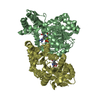
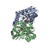
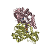
- PDB-5bk7: The structure of MppP E15A mutant soaked with the substrate L-arginine -
+ Open data

- Basic information
| Entry | Database: PDB / ID: 5bk7 | ||||||
|---|---|---|---|---|---|---|---|
| Title | The structure of MppP E15A mutant soaked with the substrate L-arginine | ||||||
 Components Components | PLP-Dependent L-Arginine Hydroxylase MppP | ||||||
 Keywords Keywords | OXIDOREDUCTASE / dimer / substrate binding in E15A mutant / oxidase / PLP / hydrolase-antibiotic complex | ||||||
| Function / homology |  Function and homology information Function and homology informationamino acid metabolic process / transaminase activity / pyridoxal phosphate binding / metal ion binding Similarity search - Function | ||||||
| Biological species |  Streptomyces wadayamensis (bacteria) Streptomyces wadayamensis (bacteria) | ||||||
| Method |  X-RAY DIFFRACTION / SYNCHROTRON / MOLECULAR REPLACEMENT / Resolution: 2.196 Å X-RAY DIFFRACTION / SYNCHROTRON / MOLECULAR REPLACEMENT / Resolution: 2.196 Å | ||||||
 Authors Authors | Han, L. / Silvaggi, N.R. | ||||||
| Funding support |  United States, 1items United States, 1items
| ||||||
 Citation Citation | Journal: Biochemistry / Year: 2018 Title: Streptomyces wadayamensis MppP is a PLP-Dependent Oxidase, Not an Oxygenase. Authors: Han, L. / Vuksanovic, N. / Oehm, S.A. / Fenske, T.G. / Schwabacher, A.W. / Silvaggi, N.R. | ||||||
| History |
|
- Structure visualization
| Structure viewer | Molecule: MolmilJmol/JSmol |
|---|
- Downloads & links
-Download
| PDBx/mmCIF format | 5bk7.cif.gz | 1 MB | Display | PDBx/mmCIF format |
|---|---|---|---|---|
| PDB format | pdb5bk7.ent.gz | 896.2 KB | Display | PDB format |
| PDBx/mmJSON format | 5bk7.json.gz | Tree view | PDBx/mmJSON format | |
| Others |  Other downloads Other downloads |
-Validation report
| Arichive directory | https://data.pdbj.org/pub/pdb/validation_reports/bk/5bk7ftp://data.pdbj.org/pub/pdb/validation_reports/bk/5bk7 | HTTPS FTP |
|---|
-Related structure data
| Related structure data |  6c8tC  6c92C  6c9bC  5dj1S S: Starting model for refinement C: citing same article ( |
|---|---|
| Similar structure data |


-Links
 PDBj
PDBj- Assembly
| Deposited unit | 
| ||||||||
|---|---|---|---|---|---|---|---|---|---|
| 1 | 
| ||||||||
| 2 | 
| ||||||||
| 3 | 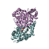
| ||||||||
| 4 | 
| ||||||||
| Unit cell |
|
-Components
| #1: Protein | Mass: 41479.719 Da / Num. of mol.: 8 / Mutation: E15A Source method: isolated from a genetically manipulated source Source: (gene. exp.) Streptomyces wadayamensis (bacteria) / Plasmid: pSUMO / Production host: #2: Chemical | ChemComp-EQJ / (   Mass: 403.328 Da / Num. of mol.: 8 / Source method: obtained synthetically / Formula: C14H22N5O7P Mass: 403.328 Da / Num. of mol.: 8 / Source method: obtained synthetically / Formula: C14H22N5O7P#3: Water | ChemComp-HOH / |  Mass: 18.015 Da / Num. of mol.: 846 / Source method: isolated from a natural source / Formula: H2O Mass: 18.015 Da / Num. of mol.: 846 / Source method: isolated from a natural source / Formula: H2O |
|---|
-Experimental details
-Experiment
| Experiment | Method: X-RAY DIFFRACTION / Number of used crystals: 1 |
|---|
- Sample preparation
| Crystal | Density Matthews: 2.65 Å3/Da / Density % sol: 53.62 % |
|---|---|
| Crystal grow | Temperature: 295 K / Method: vapor diffusion, hanging drop / pH: 6.4 Details: 0.04 M citric acid, 0.06 M BIS-TRIS Propane, 20% PEG 3350 |
-Data collection
| Diffraction | Mean temperature: 100 K |
|---|---|
| Diffraction source | Source: SYNCHROTRON / Site: APS / Beamline: 21-ID-D / Wavelength: 0.978 Å |
| Detector | Type: DECTRIS EIGER X 9M / Detector: PIXEL / Date: Oct 18, 2016 |
| Radiation | Protocol: SINGLE WAVELENGTH / Monochromatic (M) / Laue (L): M / Scattering type: x-ray |
| Radiation wavelength | Wavelength: 0.978 Å / Relative weight: 1 |
| Reflection | Resolution: 2.2→50 Å / Num. obs: 170041 / % possible obs: 97 % / Redundancy: 5.7 % / Rmerge(I) obs: 0.11 / Net I/σ(I): 14.9 |
| Reflection shell | Resolution: 2.2→2.24 Å / Redundancy: 4.5 % / Rmerge(I) obs: 0.662 / Mean I/σ(I) obs: 1.7 / Num. unique obs: 7831 / % possible all: 89.4 |
- Processing
| Software |
| |||||||||||||||||||||||||||||||||||||||||||||||||||||||||||||||||||||||||||||||||||||||||||||||||||||||||
|---|---|---|---|---|---|---|---|---|---|---|---|---|---|---|---|---|---|---|---|---|---|---|---|---|---|---|---|---|---|---|---|---|---|---|---|---|---|---|---|---|---|---|---|---|---|---|---|---|---|---|---|---|---|---|---|---|---|---|---|---|---|---|---|---|---|---|---|---|---|---|---|---|---|---|---|---|---|---|---|---|---|---|---|---|---|---|---|---|---|---|---|---|---|---|---|---|---|---|---|---|---|---|---|---|---|---|
| Refinement | Method to determine structure: MOLECULAR REPLACEMENT Starting model: 5DJ1 Resolution: 2.196→45.401 Å / SU ML: 0.3 / Cross valid method: FREE R-VALUE / σ(F): 1.36 / Phase error: 30.64 / Stereochemistry target values: ML
| |||||||||||||||||||||||||||||||||||||||||||||||||||||||||||||||||||||||||||||||||||||||||||||||||||||||||
| Solvent computation | Shrinkage radii: 0.9 Å / VDW probe radii: 1.11 Å / Solvent model: FLAT BULK SOLVENT MODEL | |||||||||||||||||||||||||||||||||||||||||||||||||||||||||||||||||||||||||||||||||||||||||||||||||||||||||
| Refinement step | Cycle: LAST / Resolution: 2.196→45.401 Å
| |||||||||||||||||||||||||||||||||||||||||||||||||||||||||||||||||||||||||||||||||||||||||||||||||||||||||
| Refine LS restraints |
| |||||||||||||||||||||||||||||||||||||||||||||||||||||||||||||||||||||||||||||||||||||||||||||||||||||||||
| LS refinement shell |
|
