+ Open data
- Basic information
| Entry |  | |||||||||
|---|---|---|---|---|---|---|---|---|---|---|
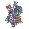
| Title | Cryo-EM structure of human PRDX4 | |||||||||
 Map data Map data | Cryo-EM structure of human PRDX4 | |||||||||
 Sample Sample |
| |||||||||
 Keywords Keywords | OXIDOREDUCTASE | |||||||||
| Function / homology |  Function and homology information Function and homology informationI-kappaB phosphorylation / negative regulation of male germ cell proliferation / thioredoxin-dependent peroxiredoxin / thioredoxin peroxidase activity / molecular sequestering activity / extracellular matrix organization / cell redox homeostasis / protein maturation / hydrogen peroxide catabolic process / male gonad development ...I-kappaB phosphorylation / negative regulation of male germ cell proliferation / thioredoxin-dependent peroxiredoxin / thioredoxin peroxidase activity / molecular sequestering activity / extracellular matrix organization / cell redox homeostasis / protein maturation / hydrogen peroxide catabolic process / male gonad development / response to oxidative stress / spermatogenesis / secretory granule lumen / molecular adaptor activity / ficolin-1-rich granule lumen / Neutrophil degranulation / endoplasmic reticulum / extracellular exosome / extracellular region / identical protein binding / nucleus / cytosol Similarity search - Function | |||||||||
| Biological species |  Homo sapiens (human) Homo sapiens (human) | |||||||||
| Method | single particle reconstruction / cryo EM / Resolution: 2.83 Å | |||||||||
 Authors Authors | Su CC / Lyu M | |||||||||
| Funding support |  United States, 1 items United States, 1 items
| |||||||||
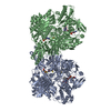
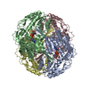
 Citation Citation |  Journal: Cell Rep / Year: 2023 Journal: Cell Rep / Year: 2023Title: High-resolution structural-omics of human liver enzymes. Authors: Chih-Chia Su / Meinan Lyu / Zhemin Zhang / Masaru Miyagi / Wei Huang / Derek J Taylor / Edward W Yu /  Abstract: We applied raw human liver microsome lysate to a holey carbon grid and used cryo-electron microscopy (cryo-EM) to define its composition. From this sample we identified and simultaneously determined ...We applied raw human liver microsome lysate to a holey carbon grid and used cryo-electron microscopy (cryo-EM) to define its composition. From this sample we identified and simultaneously determined high-resolution structural information for ten unique human liver enzymes involved in diverse cellular processes. Notably, we determined the structure of the endoplasmic bifunctional protein H6PD, where the N- and C-terminal domains independently possess glucose-6-phosphate dehydrogenase and 6-phosphogluconolactonase enzymatic activity, respectively. We also obtained the structure of heterodimeric human GANAB, an ER glycoprotein quality-control machinery that contains a catalytic α subunit and a noncatalytic β subunit. In addition, we observed a decameric peroxidase, PRDX4, which directly contacts a disulfide isomerase-related protein, ERp46. Structural data suggest that several glycosylations, bound endogenous compounds, and ions associate with these human liver enzymes. These results highlight the importance of cryo-EM in facilitating the elucidation of human organ proteomics at the atomic level. | |||||||||
| History |
|
- Structure visualization
| Supplemental images |
|---|
- Downloads & links
-EMDB archive
| Map data |  emd_28214.map.gz emd_28214.map.gz | 97.2 MB |  EMDB map data format EMDB map data format | |
|---|---|---|---|---|
| Header (meta data) |  emd-28214-v30.xml emd-28214-v30.xml emd-28214.xml emd-28214.xml | 16.2 KB 16.2 KB | Display Display |  EMDB header EMDB header |
| FSC (resolution estimation) |  emd_28214_fsc.xml emd_28214_fsc.xml emd_28214_fsc_2.xml emd_28214_fsc_2.xml | 9.8 KB 13 KB | Display Display |  FSC data file FSC data file |
| Images |  emd_28214.png emd_28214.png | 152.3 KB | ||
| Masks |  emd_28214_msk_1.map emd_28214_msk_1.map | 103 MB |  Mask map Mask map | |
| Filedesc metadata |  emd-28214.cif.gz emd-28214.cif.gz | 5.2 KB | ||
| Others |  emd_28214_additional_1.map.gz emd_28214_additional_1.map.gz emd_28214_half_map_1.map.gz emd_28214_half_map_1.map.gz emd_28214_half_map_2.map.gz emd_28214_half_map_2.map.gz | 51.8 MB 95.7 MB 95.7 MB | ||
| Archive directory |  http://ftp.pdbj.org/pub/emdb/structures/EMD-28214 http://ftp.pdbj.org/pub/emdb/structures/EMD-28214 ftp://ftp.pdbj.org/pub/emdb/structures/EMD-28214 ftp://ftp.pdbj.org/pub/emdb/structures/EMD-28214 | HTTPS FTP |
-Validation report
| Summary document |  emd_28214_validation.pdf.gz emd_28214_validation.pdf.gz | 955.1 KB | Display |  EMDB validaton report EMDB validaton report |
|---|---|---|---|---|
| Full document |  emd_28214_full_validation.pdf.gz emd_28214_full_validation.pdf.gz | 954.6 KB | Display | |
| Data in XML |  emd_28214_validation.xml.gz emd_28214_validation.xml.gz | 18.2 KB | Display | |
| Data in CIF |  emd_28214_validation.cif.gz emd_28214_validation.cif.gz | 23.5 KB | Display | |
| Arichive directory |  https://ftp.pdbj.org/pub/emdb/validation_reports/EMD-28214 https://ftp.pdbj.org/pub/emdb/validation_reports/EMD-28214 ftp://ftp.pdbj.org/pub/emdb/validation_reports/EMD-28214 ftp://ftp.pdbj.org/pub/emdb/validation_reports/EMD-28214 | HTTPS FTP |
-Related structure data
| Related structure data |  8ekwMC  7uzmC  8ekyC  8em2C  8emrC  8emsC  8emtC  8eneC  8eojC  8eorC  23432 M: atomic model generated by this map C: citing same article ( |
|---|---|
| Similar structure data | Similarity search - Function & homology  F&H Search F&H Search |
- Links
Links
| EMDB pages |  EMDB (EBI/PDBe) / EMDB (EBI/PDBe) /  EMDataResource EMDataResource |
|---|
- Map
Map
| File |  Download / File: emd_28214.map.gz / Format: CCP4 / Size: 103 MB / Type: IMAGE STORED AS FLOATING POINT NUMBER (4 BYTES) Download / File: emd_28214.map.gz / Format: CCP4 / Size: 103 MB / Type: IMAGE STORED AS FLOATING POINT NUMBER (4 BYTES) | ||||||||||||||||||||||||||||||||||||
|---|---|---|---|---|---|---|---|---|---|---|---|---|---|---|---|---|---|---|---|---|---|---|---|---|---|---|---|---|---|---|---|---|---|---|---|---|---|
| Annotation | Cryo-EM structure of human PRDX4 | ||||||||||||||||||||||||||||||||||||
| Projections & slices | Image control
Images are generated by Spider. | ||||||||||||||||||||||||||||||||||||
| Voxel size | X=Y=Z: 1.08 Å | ||||||||||||||||||||||||||||||||||||
| Density |
| ||||||||||||||||||||||||||||||||||||
| Symmetry | Space group: 1 | ||||||||||||||||||||||||||||||||||||
| Details | EMDB XML:
|
-Supplemental data
-Mask #1
| File |  emd_28214_msk_1.map emd_28214_msk_1.map | ||||||||||||
|---|---|---|---|---|---|---|---|---|---|---|---|---|---|
| Projections & Slices |
| ||||||||||||
| Density Histograms |
-Additional map: unsharpened map
| File | emd_28214_additional_1.map | ||||||||||||
|---|---|---|---|---|---|---|---|---|---|---|---|---|---|
| Annotation | unsharpened map | ||||||||||||
| Projections & Slices |
| ||||||||||||
| Density Histograms |
-Half map: Cryo-EM structure of human PRDX4
| File | emd_28214_half_map_1.map | ||||||||||||
|---|---|---|---|---|---|---|---|---|---|---|---|---|---|
| Annotation | Cryo-EM structure of human PRDX4 | ||||||||||||
| Projections & Slices |
| ||||||||||||
| Density Histograms |
-Half map: Cryo-EM structure of human PRDX4
| File | emd_28214_half_map_2.map | ||||||||||||
|---|---|---|---|---|---|---|---|---|---|---|---|---|---|
| Annotation | Cryo-EM structure of human PRDX4 | ||||||||||||
| Projections & Slices |
| ||||||||||||
| Density Histograms |
- Sample components
-Entire : H6PD
| Entire | Name: H6PD |
|---|---|
| Components |
|
-Supramolecule #1: H6PD
| Supramolecule | Name: H6PD / type: complex / ID: 1 / Parent: 0 / Macromolecule list: #1 |
|---|---|
| Source (natural) | Organism:  Homo sapiens (human) Homo sapiens (human) |
-Macromolecule #1: Peroxiredoxin-4
| Macromolecule | Name: Peroxiredoxin-4 / type: protein_or_peptide / ID: 1 / Number of copies: 10 / Enantiomer: LEVO |
|---|---|
| Source (natural) | Organism:  Homo sapiens (human) Homo sapiens (human) |
| Molecular weight | Theoretical: 30.578873 KDa |
| Sequence | String: MEALPLLAAT TPDHGRHRRL LLLPLLLFLL PAGAVQGWET EERPRTREEE CHFYAGGQVY PGEASRVSVA DHSLHLSKAK ISKPAPYWE GTAVIDGEFK ELKLTDYRGK YLVFFFYPLD FTFVCPTEII AFGDRLEEFR SINTEVVACS VDSQFTHLAW I NTPRRQGG ...String: MEALPLLAAT TPDHGRHRRL LLLPLLLFLL PAGAVQGWET EERPRTREEE CHFYAGGQVY PGEASRVSVA DHSLHLSKAK ISKPAPYWE GTAVIDGEFK ELKLTDYRGK YLVFFFYPLD FTFVCPTEII AFGDRLEEFR SINTEVVACS VDSQFTHLAW I NTPRRQGG LGPIRIPLLS DLTHQISKDY GVYLEDSGHT LRGLFIIDDK GILRQITLND LPVGRSVDET LRLVQAFQYT DK HGEVCPA GWKPGSETII PDPAGKLKYF DKLN UniProtKB: Peroxiredoxin-4 |
-Macromolecule #2: water
| Macromolecule | Name: water / type: ligand / ID: 2 / Number of copies: 402 / Formula: HOH |
|---|---|
| Molecular weight | Theoretical: 18.015 Da |
| Chemical component information |  ChemComp-HOH: |
-Experimental details
-Structure determination
| Method | cryo EM |
|---|---|
 Processing Processing | single particle reconstruction |
| Aggregation state | particle |
- Sample preparation
| Concentration | 0.5 mg/mL |
|---|---|
| Buffer | pH: 7.5 |
| Vitrification | Cryogen name: ETHANE / Chamber humidity: 100 % / Chamber temperature: 277 K / Instrument: FEI VITROBOT MARK IV |
| Details | This is from a heterogeneous and impure protein sample. |
- Electron microscopy
| Microscope | FEI TITAN KRIOS |
|---|---|
| Image recording | Film or detector model: GATAN K3 BIOQUANTUM (6k x 4k) / Average electron dose: 29.0 e/Å2 |
| Electron beam | Acceleration voltage: 300 kV / Electron source:  FIELD EMISSION GUN FIELD EMISSION GUN |
| Electron optics | Illumination mode: SPOT SCAN / Imaging mode: BRIGHT FIELD / Nominal defocus max: 2.5 µm / Nominal defocus min: 1.0 µm |
| Experimental equipment |  Model: Titan Krios / Image courtesy: FEI Company |
+ Image processing
-Atomic model buiding 1
| Refinement | Protocol: AB INITIO MODEL |
|---|---|
| Output model |  PDB-8ekw: |
 Movie
Movie Controller


 Z (Sec.)
 Y (Row.)
 X (Col.)


