Movie
Movie Controller


+ Open data

- Basic information
| Entry |  | |||||||||
|---|---|---|---|---|---|---|---|---|---|---|
| Title | Cryo-EM structure of human liver glycogen phosphorylase | |||||||||
 Map data Map data | ||||||||||
 Sample Sample |
| |||||||||
 Keywords Keywords | Glycogen Phosphorylase / PLP / HYDROLASE | |||||||||
| Function / homology |  Function and homology information Function and homology informationpurine nucleobase binding / vitamin binding / D-glucose binding / bile acid binding / glycogen phosphorylase / glycogen phosphorylase activity / glycogen catabolic process / glycogen metabolic process / Glycogen breakdown (glycogenolysis) / AMP binding ...purine nucleobase binding / vitamin binding / D-glucose binding / bile acid binding / glycogen phosphorylase / glycogen phosphorylase activity / glycogen catabolic process / glycogen metabolic process / Glycogen breakdown (glycogenolysis) / AMP binding / necroptotic process / response to bacterium / pyridoxal phosphate binding / glucose homeostasis / secretory granule lumen / ficolin-1-rich granule lumen / Neutrophil degranulation / extracellular exosome / extracellular region / ATP binding / identical protein binding / cytoplasm / cytosol Similarity search - Function | |||||||||
| Biological species |  Homo sapiens (human) Homo sapiens (human) | |||||||||
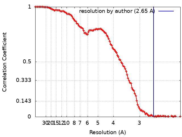
| Method | single particle reconstruction / cryo EM / Resolution: 2.65 Å | |||||||||
 Authors Authors | Su C / Lyu M / Zhang Z / Yu EW | |||||||||
| Funding support |  United States, 1 items United States, 1 items
| |||||||||
 Citation Citation | Journal: Cell Rep / Year: 2023 Title: High-resolution structural-omics of human liver enzymes. Authors: Chih-Chia Su / Meinan Lyu / Zhemin Zhang / Masaru Miyagi / Wei Huang / Derek J Taylor / Edward W Yu / Abstract: We applied raw human liver microsome lysate to a holey carbon grid and used cryo-electron microscopy (cryo-EM) to define its composition. From this sample we identified and simultaneously determined ...We applied raw human liver microsome lysate to a holey carbon grid and used cryo-electron microscopy (cryo-EM) to define its composition. From this sample we identified and simultaneously determined high-resolution structural information for ten unique human liver enzymes involved in diverse cellular processes. Notably, we determined the structure of the endoplasmic bifunctional protein H6PD, where the N- and C-terminal domains independently possess glucose-6-phosphate dehydrogenase and 6-phosphogluconolactonase enzymatic activity, respectively. We also obtained the structure of heterodimeric human GANAB, an ER glycoprotein quality-control machinery that contains a catalytic α subunit and a noncatalytic β subunit. In addition, we observed a decameric peroxidase, PRDX4, which directly contacts a disulfide isomerase-related protein, ERp46. Structural data suggest that several glycosylations, bound endogenous compounds, and ions associate with these human liver enzymes. These results highlight the importance of cryo-EM in facilitating the elucidation of human organ proteomics at the atomic level. | |||||||||
| History |
|
- Structure visualization
| Supplemental images |
|---|

- Downloads & links
-EMDB archive
| Map data | emd_28263.map.gz | 97.3 MB | EMDB map data format | |
|---|---|---|---|---|
| Header (meta data) | emd-28263-v30.xmlemd-28263.xml | 14.1 KB 14.1 KB | Display Display | EMDB header |
| FSC (resolution estimation) | emd_28263_fsc.xml | 13.7 KB | Display | FSC data file |
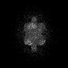
| Images |  emd_28263.png emd_28263.png | 132.8 KB | ||
| Masks | emd_28263_msk_1.map | 103 MB | Mask map | |
| Filedesc metadata | emd-28263.cif.gz | 5.5 KB | ||
| Others | emd_28263_half_map_1.map.gzemd_28263_half_map_2.map.gz | 95.4 MB 95.4 MB | ||
| Archive directory |  http://ftp.pdbj.org/pub/emdb/structures/EMD-28263ftp://ftp.pdbj.org/pub/emdb/structures/EMD-28263 http://ftp.pdbj.org/pub/emdb/structures/EMD-28263ftp://ftp.pdbj.org/pub/emdb/structures/EMD-28263 | HTTPS FTP |
-Related structure data
| Related structure data |  8emsMC 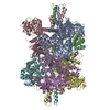 7uzmC  8ekwC  8ekyC  8em2C  8emrC 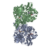 8emtC 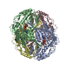 8eneC  8eojC  8eorC  23433 M: atomic model generated by this map C: citing same article ( |
|---|---|
| Similar structure data |


-Links
| EMDB pages | EMDB (EBI/PDBe) / EMDataResource |
|---|---|
| Related items in Molecule of the Month |


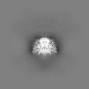
-Map
| File | Download / File: emd_28263.map.gz / Format: CCP4 / Size: 103 MB / Type: IMAGE STORED AS FLOATING POINT NUMBER (4 BYTES) | ||||||||||||||||||||||||||||||||||||
|---|---|---|---|---|---|---|---|---|---|---|---|---|---|---|---|---|---|---|---|---|---|---|---|---|---|---|---|---|---|---|---|---|---|---|---|---|---|
| Projections & slices | Image control
Images are generated by Spider. | ||||||||||||||||||||||||||||||||||||
| Voxel size | X=Y=Z: 1.08 Å | ||||||||||||||||||||||||||||||||||||
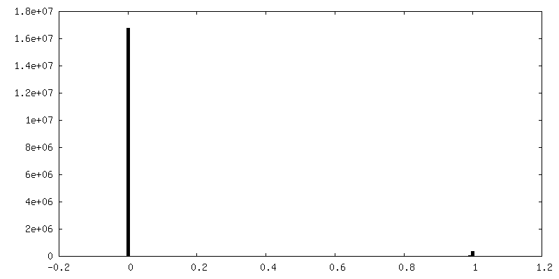
| Density |
| ||||||||||||||||||||||||||||||||||||
| Symmetry | Space group: 1 | ||||||||||||||||||||||||||||||||||||
| Details | EMDB XML:
|
 Z (Sec.)
 Y (Row.)
 X (Col.)


-Supplemental data
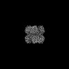
-Mask #1
| File | emd_28263_msk_1.map | ||||||||||||
|---|---|---|---|---|---|---|---|---|---|---|---|---|---|
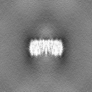
| Projections & Slices |
| ||||||||||||
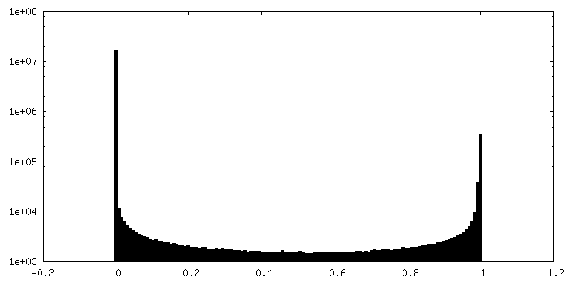
| Density Histograms |


-Half map: #2
| File | emd_28263_half_map_1.map | ||||||||||||
|---|---|---|---|---|---|---|---|---|---|---|---|---|---|
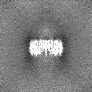
| Projections & Slices |
| ||||||||||||
| Density Histograms |


-Half map: #1
| File | emd_28263_half_map_2.map | ||||||||||||
|---|---|---|---|---|---|---|---|---|---|---|---|---|---|
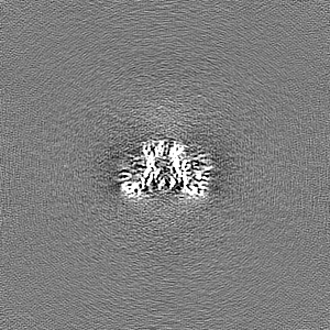
| Projections & Slices |
| ||||||||||||
| Density Histograms |


- Sample components
-Entire : Glycogen Phosphorylase
| Entire | Name: Glycogen Phosphorylase |
|---|---|
| Components |
|
-Supramolecule #1: Glycogen Phosphorylase
| Supramolecule | Name: Glycogen Phosphorylase / type: complex / ID: 1 / Parent: 0 / Macromolecule list: #1 |
|---|---|
| Source (natural) | Organism: Homo sapiens (human) |
-Macromolecule #1: Glycogen phosphorylase, liver form
| Macromolecule | Name: Glycogen phosphorylase, liver form / type: protein_or_peptide / ID: 1 / Number of copies: 2 / Enantiomer: LEVO / EC number: glycogen phosphorylase |
|---|---|
| Source (natural) | Organism: Homo sapiens (human) / Organ: liver |
| Molecular weight | Theoretical: 95.376406 KDa |
| Sequence | String: MAKPLTDQEK RRQISIRGIV GVENVAELKK SFNRHLHFTL VKDRNVATTR DYYFALAHTV RDHLVGRWIR TQQHYYDKCP KRVYYLSLE FYMGRTLQNT MINLGLQNAC DEAIYQLGLD IEELEEIEED AGLGNGGLGR LAACFLDSMA TLGLAAYGYG I RYEYGIFN ...String: MAKPLTDQEK RRQISIRGIV GVENVAELKK SFNRHLHFTL VKDRNVATTR DYYFALAHTV RDHLVGRWIR TQQHYYDKCP KRVYYLSLE FYMGRTLQNT MINLGLQNAC DEAIYQLGLD IEELEEIEED AGLGNGGLGR LAACFLDSMA TLGLAAYGYG I RYEYGIFN QKIRDGWQVE EADDWLRYGN PWEKSRPEFM LPVHFYGKVE HTNTGTKWID TQVVLALPYD TPVPGYMNNT VN TMRLWSA RAPNDFNLRD FNVGDYIQAV LDRNLAENIS RVLYPNDNFF EGKELRLKQE YFVVAATLQD IIRRFKASKF GST RGAGTV FDAFPDQVAI QLNDTHPALA IPELMRIFVD IEKLPWSKAW ELTQKTFAYT NHTVLPEALE RWPVDLVEKL LPRH LEIIY EINQKHLDRI VALFPKDVDR LRRMSLIEEE GSKRINMAHL CIVGSHAVNG VAKIHSDIVK TKVFKDFSEL EPDKF QNKT NGITPRRWLL LCNPGLAELI AEKIGEDYVK DLSQLTKLHS FLGDDVFLRE LAKVKQENKL KFSQFLETEY KVKINP SSM FDVQVKRIHE YKRQLLNCLH VITMYNRIKK DPKKLFVPRT VIIGGKAAPG YHMAKMIIKL ITSVADVVNN DPMVGSK LK VIFLENYRVS LAEKVIPATD LSEQISTAGT EASGTGNMKF MLNGALTIGT MDGANVEMAE EAGEENLFIF GMRIDDVA A LDKKGYEAKE YYEALPELKL VIDQIDNGFF SPKQPDLFKD IINMLFYHDR FKVFADYEAY VKCQDKVSQL YMNPKAWNT MVLKNIAASG KFSSDRTIKE YAQNIWNVE UniProtKB: Glycogen phosphorylase, liver form |
-Macromolecule #2: PYRIDOXAL-5'-PHOSPHATE
| Macromolecule | Name: PYRIDOXAL-5'-PHOSPHATE / type: ligand / ID: 2 / Number of copies: 2 / Formula: PLP |
|---|---|
| Molecular weight | Theoretical: 247.142 Da |
| Chemical component information |  ChemComp-PLP: |
-Experimental details
-Structure determination
| Method | cryo EM |
|---|---|
 Processing Processing | single particle reconstruction |
| Aggregation state | particle |
-Sample preparation
| Buffer | pH: 7.5 |
|---|---|
| Vitrification | Cryogen name: ETHANE |
- Electron microscopy
| Microscope | FEI TITAN KRIOS |
|---|---|
| Image recording | Film or detector model: GATAN K3 BIOQUANTUM (6k x 4k) / Average electron dose: 41.25 e/Å2 |
| Electron beam | Acceleration voltage: 300 kV / Electron source:  FIELD EMISSION GUN FIELD EMISSION GUN |
| Electron optics | Illumination mode: FLOOD BEAM / Imaging mode: BRIGHT FIELD / Nominal defocus max: 2.5 µm / Nominal defocus min: 1.0 µm |
| Experimental equipment |  Model: Titan Krios / Image courtesy: FEI Company |