 Movie
Movie Controller

[English] 日本語
 Yorodumi
- EMDB-25563: Cryo-EM structure of the extracellular module of the full-length ... -
+ Open data

- Basic information
| Entry | Database: EMDB / ID: EMD-25563 | |||||||||
|---|---|---|---|---|---|---|---|---|---|---|
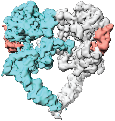
| Title | Cryo-EM structure of the extracellular module of the full-length EGFR bound to TGF-alpha. "tips-juxtaposed" conformation | |||||||||
 Map data Map data | WT:TGFa_jux | |||||||||
 Sample Sample |
| |||||||||
 Keywords Keywords | receptor tyrosine kinases / epidermal growth factor receptor / SIGNALING PROTEIN / Transferase | |||||||||
| Function / homology |  Function and homology information Function and homology informationhepatocyte proliferation / transmembrane receptor protein tyrosine kinase activator activity / Cargo concentration in the ER / COPII-mediated vesicle transport / multivesicular body, internal vesicle lumen / negative regulation of cardiocyte differentiation / Shc-EGFR complex / morphogenesis of an epithelial fold / positive regulation of protein kinase C signaling / Inhibition of Signaling by Overexpressed EGFR ...hepatocyte proliferation / transmembrane receptor protein tyrosine kinase activator activity / Cargo concentration in the ER / COPII-mediated vesicle transport / multivesicular body, internal vesicle lumen / negative regulation of cardiocyte differentiation / Shc-EGFR complex / morphogenesis of an epithelial fold / positive regulation of protein kinase C signaling / Inhibition of Signaling by Overexpressed EGFR / epidermal growth factor receptor activity / EGFR interacts with phospholipase C-gamma / epidermal growth factor binding / epidermal growth factor receptor binding / response to UV-A / regulation of peptidyl-tyrosine phosphorylation / ubiquitin-dependent endocytosis / PLCG1 events in ERBB2 signaling / digestive tract morphogenesis / ERBB2-EGFR signaling pathway / PTK6 promotes HIF1A stabilization / eyelid development in camera-type eye / ERBB2 Activates PTK6 Signaling / Signaling by EGFR / intracellular vesicle / cerebral cortex cell migration / hair follicle development / ERBB2 Regulates Cell Motility / Developmental Lineage of Mammary Gland Myoepithelial Cells / mammary gland alveolus development / protein insertion into membrane / positive regulation of cell division / protein tyrosine kinase activator activity / Signaling by ERBB4 / Respiratory syncytial virus (RSV) attachment and entry / embryonic placenta development / negative regulation of epidermal growth factor receptor signaling pathway / PI3K events in ERBB2 signaling / positive regulation of phosphorylation / positive regulation of peptidyl-serine phosphorylation / MAP kinase kinase kinase activity / Estrogen-dependent nuclear events downstream of ESR-membrane signaling / GAB1 signalosome / salivary gland morphogenesis / xenobiotic transport / positive regulation of G1/S transition of mitotic cell cycle / positive regulation of epidermal growth factor receptor signaling pathway / epithelial cell proliferation / Signaling by ERBB2 / TFAP2 (AP-2) family regulates transcription of growth factors and their receptors / positive regulation of mitotic nuclear division / transmembrane receptor protein tyrosine kinase activity / endoplasmic reticulum-Golgi intermediate compartment membrane / GRB2 events in EGFR signaling / SHC1 events in EGFR signaling / EGFR Transactivation by Gastrin / positive regulation of epithelial cell proliferation / GRB2 events in ERBB2 signaling / SHC1 events in ERBB2 signaling / ossification / cellular response to epidermal growth factor stimulus / basal plasma membrane / positive regulation of DNA replication / positive regulation of DNA repair / Signal transduction by L1 / growth factor activity / positive regulation of protein localization to plasma membrane / cellular response to amino acid stimulus / cellular response to estradiol stimulus / phosphatidylinositol 3-kinase/protein kinase B signal transduction / sperm end piece / NOTCH3 Activation and Transmission of Signal to the Nucleus / ER to Golgi transport vesicle membrane / clathrin-coated endocytic vesicle membrane / sperm principal piece / Signaling by ERBB2 TMD/JMD mutants / cell-cell adhesion / EGFR downregulation / Constitutive Signaling by EGFRvIII / receptor protein-tyrosine kinase / negative regulation of protein catabolic process / Signaling by ERBB2 ECD mutants / Signaling by ERBB2 KD Mutants / positive regulation of protein phosphorylation / positive regulation of miRNA transcription / cell morphogenesis / positive regulation of fibroblast proliferation / epidermal growth factor receptor signaling pathway / ruffle membrane / kinase binding / Downregulation of ERBB2 signaling / neuron differentiation / Constitutive Signaling by Aberrant PI3K in Cancer / HCMV Early Events / cell junction / positive regulation of canonical Wnt signaling pathway / actin filament binding / transmembrane signaling receptor activity / PIP3 activates AKT signaling / Cargo recognition for clathrin-mediated endocytosis Similarity search - Function | |||||||||
| Biological species |  Homo sapiens (human) Homo sapiens (human) | |||||||||
| Method | single particle reconstruction / cryo EM / Resolution: 3.4 Å | |||||||||
 Authors Authors | Huang Y / Ognjenovic J | |||||||||
| Funding support |  Canada, Canada,  United States, 2 items United States, 2 items
| |||||||||
 Citation Citation | Journal: Elife / Year: 2021 Title: A molecular mechanism for the generation of ligand-dependent differential outputs by the epidermal growth factor receptor. Authors: Yongjian Huang / Jana Ognjenovic / Deepti Karandur / Kate Miller / Alan Merk / Sriram Subramaniam / John Kuriyan / Abstract: The epidermal growth factor receptor (EGFR) is a receptor tyrosine kinase that couples the binding of extracellular ligands, such as EGF and transforming growth factor-α (TGF-α), to the initiation ...The epidermal growth factor receptor (EGFR) is a receptor tyrosine kinase that couples the binding of extracellular ligands, such as EGF and transforming growth factor-α (TGF-α), to the initiation of intracellular signaling pathways. EGFR binds to EGF and TGF-α with similar affinity, but generates different signals from these ligands. To address the mechanistic basis of this phenomenon, we have carried out cryo-EM analyses of human EGFR bound to EGF and TGF-α. We show that the extracellular module adopts an ensemble of dimeric conformations when bound to either EGF or TGF-α. The two extreme states of this ensemble represent distinct ligand-bound quaternary structures in which the membrane-proximal tips of the extracellular module are either juxtaposed or separated. EGF and TGF-α differ in their ability to maintain the conformation with the membrane-proximal tips of the extracellular module separated, and this conformation is stabilized preferentially by an oncogenic EGFR mutation. Close proximity of the transmembrane helices at the junction with the extracellular module has been associated previously with increased EGFR activity. Our results show how EGFR can couple the binding of different ligands to differential modulation of this proximity, thereby suggesting a molecular mechanism for the generation of ligand-sensitive differential outputs in this receptor family. | |||||||||
| History |
|
- Structure visualization
| Movie |
Movie viewer |
|---|---|
| Structure viewer | EM map: SurfViewMolmilJmol/JSmol |
| Supplemental images |
- Downloads & links
-EMDB archive
| Map data | emd_25563.map.gz | 122.7 MB | EMDB map data format | |
|---|---|---|---|---|
| Header (meta data) | emd-25563-v30.xmlemd-25563.xml | 13.2 KB 13.2 KB | Display Display | EMDB header |
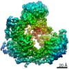
| Images |  emd_25563.png emd_25563.png | 175.9 KB | ||
| Filedesc metadata | emd-25563.cif.gz | 6.1 KB | ||
| Archive directory |  http://ftp.pdbj.org/pub/emdb/structures/EMD-25563ftp://ftp.pdbj.org/pub/emdb/structures/EMD-25563 http://ftp.pdbj.org/pub/emdb/structures/EMD-25563ftp://ftp.pdbj.org/pub/emdb/structures/EMD-25563 | HTTPS FTP |
-Related structure data
| Related structure data |  7sz7MC  7sydC  7syeC  7sz0C  7sz1C  7sz5C C: citing same article ( M: atomic model generated by this map |
|---|---|
| Similar structure data |


-Links
| EMDB pages | EMDB (EBI/PDBe) / EMDataResource |
|---|---|
| Related items in Molecule of the Month |


-Map
| File | Download / File: emd_25563.map.gz / Format: CCP4 / Size: 244.1 MB / Type: IMAGE STORED AS FLOATING POINT NUMBER (4 BYTES) | ||||||||||||||||||||||||||||||||||||||||||||||||||||||||||||
|---|---|---|---|---|---|---|---|---|---|---|---|---|---|---|---|---|---|---|---|---|---|---|---|---|---|---|---|---|---|---|---|---|---|---|---|---|---|---|---|---|---|---|---|---|---|---|---|---|---|---|---|---|---|---|---|---|---|---|---|---|---|
| Annotation | WT:TGFa_jux | ||||||||||||||||||||||||||||||||||||||||||||||||||||||||||||
| Projections & slices | Image control
Images are generated by Spider. | ||||||||||||||||||||||||||||||||||||||||||||||||||||||||||||
| Voxel size | X=Y=Z: 1.0794 Å | ||||||||||||||||||||||||||||||||||||||||||||||||||||||||||||
| Density |
| ||||||||||||||||||||||||||||||||||||||||||||||||||||||||||||
| Symmetry | Space group: 1 | ||||||||||||||||||||||||||||||||||||||||||||||||||||||||||||
| Details | EMDB XML:
CCP4 map header:
| ||||||||||||||||||||||||||||||||||||||||||||||||||||||||||||
 Z (Sec.)
 Y (Row.)
 X (Col.)


-Supplemental data
- Sample components
-Entire : full-length human EGFR:TGF-alpha complex
| Entire | Name: full-length human EGFR:TGF-alpha complex |
|---|---|
| Components |
|
-Supramolecule #1: full-length human EGFR:TGF-alpha complex
| Supramolecule | Name: full-length human EGFR:TGF-alpha complex / type: complex / ID: 1 / Parent: 0 / Macromolecule list: all |
|---|---|
| Source (natural) | Organism: Homo sapiens (human) |
-Macromolecule #1: Epidermal growth factor receptor
| Macromolecule | Name: Epidermal growth factor receptor / type: protein_or_peptide / ID: 1 / Number of copies: 2 / Enantiomer: LEVO / EC number: receptor protein-tyrosine kinase |
|---|---|
| Source (natural) | Organism: Homo sapiens (human) |
| Molecular weight | Theoretical: 134.433328 KDa |
| Recombinant expression | Organism: Homo sapiens (human) |
| Sequence | String: MRPSGTAGAA LLALLAALCP ASRALEEKKV CQGTSNKLTQ LGTFEDHFLS LQRMFNNCEV VLGNLEITYV QRNYDLSFLK TIQEVAGYV LIALNTVERI PLENLQIIRG NMYYENSYAL AVLSNYDANK TGLKELPMRN LQEILHGAVR FSNNPALCNV E SIQWRDIV ...String: MRPSGTAGAA LLALLAALCP ASRALEEKKV CQGTSNKLTQ LGTFEDHFLS LQRMFNNCEV VLGNLEITYV QRNYDLSFLK TIQEVAGYV LIALNTVERI PLENLQIIRG NMYYENSYAL AVLSNYDANK TGLKELPMRN LQEILHGAVR FSNNPALCNV E SIQWRDIV SSDFLSNMSM DFQNHLGSCQ KCDPSCPNGS CWGAGEENCQ KLTKIICAQQ CSGRCRGKSP SDCCHNQCAA GC TGPRESD CLVCRKFRNE ATCKDTCPPL MLYNPTTYQM DVNPEGKYSF GATCVKKCPR NYVVTDHGSC VRACGADSYE MEE DGVRKC KKCEGPCRKV CNGIGIGEFK DSLSINATNI KHFKNCTSIS GDLHILPVAF RGDSFTHTPP LDPQELDILK TVKE ITGFL LIQAWPENRT DLHAFENLEI IRGRTKQHGQ FSLAVVSLNI TSLGLRSLKE ISDGDVIISG NKNLCYANTI NWKKL FGTS GQKTKIISNR GENSCKATGQ VCHALCSPEG CWGPEPRDCV SCRNVSRGRE CVDKCNLLEG EPREFVENSE CIQCHP ECL PQAMNITCTG RGPDNCIQCA HYIDGPHCVK TCPAGVMGEN NTLVWKYADA GHVCHLCHPN CTYGCTGPGL EGCPTNG PK IPSIATGMVG ALLLLLVVAL GIGLFMRRRH IVRKRTLRRL LQERELVEPL TPSGEAPNQA LLRILKETEF KKIKVLGS G AFGTVYKGLW IPEGEKVKIP VAIKELREAT SPKANKEILD EAYVMASVDN PHVCRLLGIC LTSTVQLITQ LMPFGCLLD YVREHKDNIG SQYLLNWCVQ IAKGMNYLED RRLVHRDLAA RNVLVKTPQH VKITDFGLAK LLGAEEKEYH AEGGKVPIKW MALESILHR IYTHQSDVWS YGVTVWELMT FGSKPYDGIP ASEISSILEK GERLPQPPIC TIDVYMIMVK CWMIDADSRP K FRELIIEF SKMARDPQRY LVIQGDERMH LPSPTDSNFY RALMDEEDMD DVVDADEYLI PQQGFFSSPS TSRTPLLSSL SA TSNNSTV ACIDRNGLQS CPIKEDSFLQ RYSSDPTGAL TEDSIDDTFL PVPEYINQSV PKRPAGSVQN PVYHNQPLNP APS RDPHYQ DPHSTAVGNP EYLNTVQPTC VNSTFDSPAH WAQKGSHQIS LDNPDYQQDF FPKEAKPNGI FKGSTAENAE YLRV APQSS EFIGA UniProtKB: Epidermal growth factor receptor |
-Macromolecule #2: Transforming growth factor alpha
| Macromolecule | Name: Transforming growth factor alpha / type: protein_or_peptide / ID: 2 / Number of copies: 2 / Enantiomer: LEVO |
|---|---|
| Source (natural) | Organism: Homo sapiens (human) |
| Molecular weight | Theoretical: 5.560246 KDa |
| Recombinant expression | Organism:  |
| Sequence | String: VVSHFNDCPD SHTQFCFHGT CRFLVQEDKP ACVCHSGYVG ARCEHADLLA UniProtKB: Protransforming growth factor alpha |
-Experimental details
-Structure determination
| Method | cryo EM |
|---|---|
 Processing Processing | single particle reconstruction |
| Aggregation state | particle |
-Sample preparation
| Concentration | 0.5 mg/mL |
|---|---|
| Buffer | pH: 8 |
| Grid | Model: UltrAuFoil R1.2/1.3 / Pretreatment - Type: PLASMA CLEANING |
| Vitrification | Cryogen name: ETHANE |
- Electron microscopy
| Microscope | FEI TITAN KRIOS |
|---|---|
| Image recording | Film or detector model: GATAN K3 (6k x 4k) / Average electron dose: 50.0 e/Å2 |
| Electron beam | Acceleration voltage: 300 kV / Electron source:  FIELD EMISSION GUN FIELD EMISSION GUN |
| Electron optics | Illumination mode: FLOOD BEAM / Imaging mode: BRIGHT FIELD |
| Experimental equipment |  Model: Titan Krios / Image courtesy: FEI Company |
