Movie
Movie Controller

[English] 日本語
 Yorodumi
- PDB-7mn6: Structure of the HER2 S310F/HER3/NRG1b Heterodimer Extracellular ... -
+ Open data

- Basic information
| Entry | Database: PDB / ID: 7mn6 | ||||||||||||
|---|---|---|---|---|---|---|---|---|---|---|---|---|---|
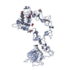
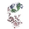
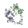
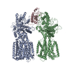
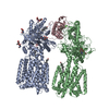
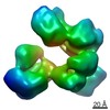
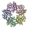
| Title | Structure of the HER2 S310F/HER3/NRG1b Heterodimer Extracellular Domain | ||||||||||||
 Components Components |
| ||||||||||||
 Keywords Keywords | SIGNALING PROTEIN / Complex / Receptor Tyrosine Kinase | ||||||||||||
| Function / homology |  Function and homology information Function and homology informationpositive regulation of cardiac muscle tissue development / neuregulin binding / cranial nerve development / Schwann cell differentiation / neuregulin receptor activity / negative regulation of secretion / endocardial cushion development / negative regulation of immature T cell proliferation in thymus / ERBB3:ERBB2 complex / ERBB2-ERBB4 signaling pathway ...positive regulation of cardiac muscle tissue development / neuregulin binding / cranial nerve development / Schwann cell differentiation / neuregulin receptor activity / negative regulation of secretion / endocardial cushion development / negative regulation of immature T cell proliferation in thymus / ERBB3:ERBB2 complex / ERBB2-ERBB4 signaling pathway / immature T cell proliferation in thymus / GRB7 events in ERBB2 signaling / RNA polymerase I core binding / semaphorin receptor complex / positive regulation of calcineurin-NFAT signaling cascade / peripheral nervous system development / Developmental Lineage of Mammary Stem Cells / negative regulation of cell adhesion / ErbB-3 class receptor binding / negative regulation of motor neuron apoptotic process / motor neuron axon guidance / Sema4D induced cell migration and growth-cone collapse / motor neuron apoptotic process / regulation of microtubule-based process / PLCG1 events in ERBB2 signaling / ERBB2-EGFR signaling pathway / enzyme-linked receptor protein signaling pathway / ERBB2 Activates PTK6 Signaling / growth factor binding / ERBB2-ERBB3 signaling pathway / neurotransmitter receptor localization to postsynaptic specialization membrane / Drug-mediated inhibition of ERBB2 signaling / Resistance of ERBB2 KD mutants to trastuzumab / Resistance of ERBB2 KD mutants to sapitinib / Resistance of ERBB2 KD mutants to tesevatinib / Resistance of ERBB2 KD mutants to neratinib / Resistance of ERBB2 KD mutants to osimertinib / Resistance of ERBB2 KD mutants to afatinib / Resistance of ERBB2 KD mutants to AEE788 / Resistance of ERBB2 KD mutants to lapatinib / Drug resistance in ERBB2 TMD/JMD mutants / positive regulation of MAP kinase activity / neuromuscular junction development / positive regulation of Rho protein signal transduction / detection of maltose stimulus / positive regulation of transcription by RNA polymerase I / maltose transport complex / ERBB2 Regulates Cell Motility / Developmental Lineage of Mammary Gland Myoepithelial Cells / oligodendrocyte differentiation / semaphorin-plexin signaling pathway / carbohydrate transport / protein tyrosine kinase activator activity / Signaling by ERBB4 / PI3K events in ERBB2 signaling / lateral plasma membrane / regulation of angiogenesis / positive regulation of protein targeting to membrane / negative regulation of signal transduction / carbohydrate transmembrane transporter activity / maltose binding / maltose transport / maltodextrin transmembrane transport / regulation of ERK1 and ERK2 cascade / Schwann cell development / coreceptor activity / ATP-binding cassette (ABC) transporter complex, substrate-binding subunit-containing / extrinsic apoptotic signaling pathway in absence of ligand / Signaling by ERBB2 / TFAP2 (AP-2) family regulates transcription of growth factors and their receptors / peptidyl-tyrosine phosphorylation / transmembrane receptor protein tyrosine kinase activity / myelination / GRB2 events in ERBB2 signaling / positive regulation of cell adhesion / ATP-binding cassette (ABC) transporter complex / cell surface receptor protein tyrosine kinase signaling pathway / SHC1 events in ERBB2 signaling / cellular response to epidermal growth factor stimulus / basal plasma membrane / Constitutive Signaling by Overexpressed ERBB2 / positive regulation of epithelial cell proliferation / Downregulation of ERBB2:ERBB3 signaling / positive regulation of translation / cell chemotaxis / neuromuscular junction / wound healing / phosphatidylinositol 3-kinase/protein kinase B signal transduction / Signaling by ERBB2 TMD/JMD mutants / receptor protein-tyrosine kinase / Signaling by ERBB2 ECD mutants / Signaling by ERBB2 KD Mutants / receptor tyrosine kinase binding / cellular response to growth factor stimulus / epidermal growth factor receptor signaling pathway / ruffle membrane / Downregulation of ERBB2 signaling / Constitutive Signaling by Aberrant PI3K in Cancer / neuron differentiation / transmembrane signaling receptor activity Similarity search - Function | ||||||||||||
| Biological species |  Homo sapiens (human) Homo sapiens (human) | ||||||||||||
| Method | ELECTRON MICROSCOPY / single particle reconstruction / cryo EM / Resolution: 3.09 Å | ||||||||||||
 Authors Authors | Diwanji, D. / Trenker, R. / Verba, K.A. / Jura, N. | ||||||||||||
| Funding support |  United States, United States,  Germany, 3items Germany, 3items
| ||||||||||||
 Citation Citation | Journal: Nature / Year: 2021 Title: Structures of the HER2-HER3-NRG1β complex reveal a dynamic dimer interface. Authors: Devan Diwanji / Raphael Trenker / Tarjani M Thaker / Feng Wang / David A Agard / Kliment A Verba / Natalia Jura / Abstract: Human epidermal growth factor receptor 2 (HER2) and HER3 form a potent pro-oncogenic heterocomplex upon binding of growth factor neuregulin-1β (NRG1β). The mechanism by which HER2 and HER3 interact ...Human epidermal growth factor receptor 2 (HER2) and HER3 form a potent pro-oncogenic heterocomplex upon binding of growth factor neuregulin-1β (NRG1β). The mechanism by which HER2 and HER3 interact remains unknown in the absence of any structures of the complex. Here we isolated the NRG1β-bound near full-length HER2-HER3 dimer and, using cryo-electron microscopy, reconstructed the extracellulardomain module, revealing unexpected dynamics at the HER2-HER3 dimerization interface. We show that the dimerization arm of NRG1β-bound HER3 is unresolved because the apo HER2 monomer does not undergo a ligand-induced conformational change needed to establish a HER3 dimerization arm-binding pocket. In a structure of the oncogenic extracellular domain mutant HER2(S310F), we observe a compensatory interaction with the HER3 dimerization arm that stabilizes the dimerization interface. Both HER2-HER3 and HER2(S310F)-HER3 retain the capacity to bind to the HER2-directed therapeutic antibody trastuzumab, but the mutant complex does not bind to pertuzumab. Our structure of the HER2(S310F)-HER3-NRG1β-trastuzumab Fab complex reveals that the receptor dimer undergoes a conformational change to accommodate trastuzumab. Thus, similar to oncogenic mutations, therapeutic agents exploit the intrinsic dynamics of the HER2-HER3 heterodimer. The unique features of a singly liganded HER2-HER3 heterodimer underscore the allosteric sensing of ligand occupancy by the dimerization interface and explain why extracellular domains of HER2 do not homo-associate via a canonical active dimer interface. | ||||||||||||
| History |
|
- Structure visualization
| Movie |
Movie viewer |
|---|---|
| Structure viewer | Molecule: MolmilJmol/JSmol |
- Downloads & links
-Download
| PDBx/mmCIF format | 7mn6.cif.gz | 285.7 KB | Display | PDBx/mmCIF format |
|---|---|---|---|---|
| PDB format | pdb7mn6.ent.gz | 209.3 KB | Display | PDB format |
| PDBx/mmJSON format | 7mn6.json.gz | Tree view | PDBx/mmJSON format | |
| Others |  Other downloads Other downloads |
-Validation report
| Arichive directory | https://data.pdbj.org/pub/pdb/validation_reports/mn/7mn6ftp://data.pdbj.org/pub/pdb/validation_reports/mn/7mn6 | HTTPS FTP |
|---|
-Related structure data
| Related structure data | 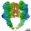 23917MC 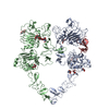 7mn5C 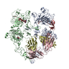 7mn8C M: map data used to model this data C: citing same article ( |
|---|---|
| Similar structure data |


-Links
 PDBj
PDBj


- Assembly
| Deposited unit | 
|
|---|---|
| 1 |
|
-Components
-Receptor tyrosine-protein kinase erbB- ... , 2 types, 2 molecules AB
| #1: Protein | Mass: 117852.719 Da / Num. of mol.: 1 / Fragment: Extracellular Domain Source method: isolated from a genetically manipulated source Source: (gene. exp.) Homo sapiens (human) / Gene: ERBB3, HER3 / Production host: Homo sapiens (human)References: UniProt: P21860, receptor protein-tyrosine kinase |
|---|---|
| #2: Protein | Mass: 160556.562 Da / Num. of mol.: 1 / Fragment: Extracellular Domain / Mutation: S310F Source method: isolated from a genetically manipulated source Details: This mutation is oncogenic Source: (gene. exp.) Homo sapiens (human), (gene. exp.) Gene: ERBB2, HER2, MLN19, NEU, NGL, malE, b4034, JW3994 / Strain: K12 / Production host: Homo sapiens (human)References: UniProt: P04626, UniProt: P0AEX9, receptor protein-tyrosine kinase |
-Protein , 1 types, 1 molecules H
| #3: Protein | Mass: 9748.859 Da / Num. of mol.: 1 / Fragment: EGF-like Domain Source method: isolated from a genetically manipulated source Source: (gene. exp.) Homo sapiens (human) / Gene: NRG1, GGF, HGL, HRGA, NDF, SMDF / Production host: |
|---|
-Sugars , 3 types, 8 molecules 
| #4: Polysaccharide | Source method: isolated from a genetically manipulated source #5: Polysaccharide | alpha-D-mannopyranose-(1-3)-beta-D-mannopyranose-(1-4)-2-acetamido-2-deoxy-beta-D-glucopyranose-(1- ...alpha-D-mannopyranose-(1-3)-beta-D-mannopyranose-(1-4)-2-acetamido-2-deoxy-beta-D-glucopyranose-(1-4)-2-acetamido-2-deoxy-beta-D-glucopyranose | Source method: isolated from a genetically manipulated source #6: Sugar | ChemComp-NAG /  Type: D-saccharide, beta linking / Mass: 221.208 Da / Num. of mol.: 5 Type: D-saccharide, beta linking / Mass: 221.208 Da / Num. of mol.: 5Source method: isolated from a genetically manipulated source Formula: C8H15NO6 |
|---|
-Details
| Has ligand of interest | N |
|---|---|
| Has protein modification | Y |
-Experimental details
-Experiment
| Experiment | Method: ELECTRON MICROSCOPY |
|---|---|
| EM experiment | Aggregation state: PARTICLE / 3D reconstruction method: single particle reconstruction |
- Sample preparation
| Component |
| ||||||||||||||||||||||||||||||
|---|---|---|---|---|---|---|---|---|---|---|---|---|---|---|---|---|---|---|---|---|---|---|---|---|---|---|---|---|---|---|---|
| Molecular weight | Experimental value: NO | ||||||||||||||||||||||||||||||
| Source (natural) |
| ||||||||||||||||||||||||||||||
| Source (recombinant) |
| ||||||||||||||||||||||||||||||
| Buffer solution | pH: 7.4 | ||||||||||||||||||||||||||||||
| Specimen | Embedding applied: NO / Shadowing applied: NO / Staining applied: NO / Vitrification applied: YES | ||||||||||||||||||||||||||||||
| Vitrification | Cryogen name: ETHANE |
- Electron microscopy imaging
| Experimental equipment |  Model: Titan Krios / Image courtesy: FEI Company |
|---|---|
| Microscopy | Model: FEI TITAN KRIOS |
| Electron gun | Electron source:  FIELD EMISSION GUN / Accelerating voltage: 300 kV / Illumination mode: FLOOD BEAM FIELD EMISSION GUN / Accelerating voltage: 300 kV / Illumination mode: FLOOD BEAM |
| Electron lens | Mode: BRIGHT FIELD |
| Image recording | Electron dose: 67 e/Å2 / Film or detector model: GATAN K3 (6k x 4k) |
- Processing
| EM software |
| ||||||||||||||||||||||||||||||||
|---|---|---|---|---|---|---|---|---|---|---|---|---|---|---|---|---|---|---|---|---|---|---|---|---|---|---|---|---|---|---|---|---|---|
| CTF correction | Type: PHASE FLIPPING AND AMPLITUDE CORRECTION | ||||||||||||||||||||||||||||||||
| 3D reconstruction | Resolution: 3.09 Å / Resolution method: FSC 0.143 CUT-OFF / Num. of particles: 99755 / Symmetry type: POINT | ||||||||||||||||||||||||||||||||
| Atomic model building |
|