- EMDB-23917: The HER2 S310F/HER3/NRG1b Heterodimer -
+
Open data
ID or keywords:
Loading...
-
Basic information
Entry
Database: EMDB / ID: EMD-23917
Title
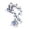
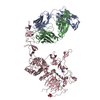
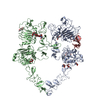
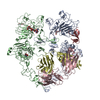
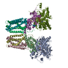
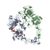
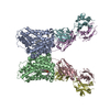
The HER2 S310F/HER3/NRG1b Heterodimer
Map data
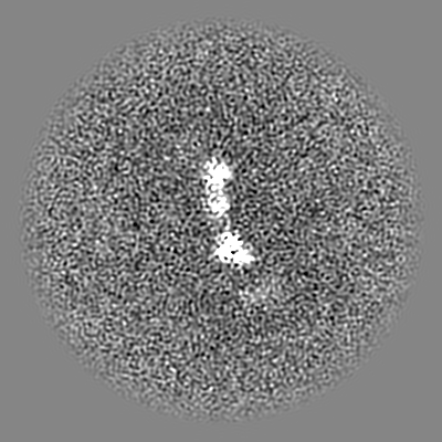
Final HER2S310F/HER3/NRG1b ectodomain reconstruction at 3.1A resolution based on 0.143FSC metric. Filtered with the FSC and sharpened with b-factor of -90.
Sample
Complex: NRB1b-bound HER2-S310F/HER3 Heterodimer in the context of near full-length receptors
Complex: HER3
Protein or peptide: Receptor tyrosine-protein kinase erbB-3
Complex: HER2-S310F, MBP Fusion
Protein or peptide: Receptor tyrosine-protein kinase erbB-2,Maltose/maltodextrin-binding periplasmic protein
Complex: Neuregulin-1
Protein or peptide: Isoform 6 of Pro-neuregulin-1, membrane-bound isoform
Ligand: 2-acetamido-2-deoxy-beta-D-glucopyranose
Keywords
Complex / Receptor Tyrosine Kinase / SIGNALING PROTEIN
Function / homology
Function and homology information
positive regulation of cardiac muscle tissue development / neuregulin binding / cranial nerve development / Schwann cell differentiation / neuregulin receptor activity / negative regulation of secretion / endocardial cushion development / negative regulation of immature T cell proliferation in thymus / ERBB3:ERBB2 complex / ERBB2-ERBB4 signaling pathway ...positive regulation of cardiac muscle tissue development / neuregulin binding / cranial nerve development / Schwann cell differentiation / neuregulin receptor activity / negative regulation of secretion / endocardial cushion development / negative regulation of immature T cell proliferation in thymus / ERBB3:ERBB2 complex / ERBB2-ERBB4 signaling pathway / immature T cell proliferation in thymus / GRB7 events in ERBB2 signaling / RNA polymerase I core binding / semaphorin receptor complex / positive regulation of calcineurin-NFAT signaling cascade / peripheral nervous system development / Developmental Lineage of Mammary Stem Cells / negative regulation of cell adhesion / ErbB-3 class receptor binding / negative regulation of motor neuron apoptotic process / Sema4D induced cell migration and growth-cone collapse / motor neuron axon guidance / regulation of microtubule-based process / motor neuron apoptotic process / PLCG1 events in ERBB2 signaling / enzyme-linked receptor protein signaling pathway / ERBB2 Activates PTK6 Signaling / growth factor binding / ERBB2-EGFR signaling pathway / ERBB2-ERBB3 signaling pathway / neurotransmitter receptor localization to postsynaptic specialization membrane / Drug-mediated inhibition of ERBB2 signaling / Resistance of ERBB2 KD mutants to trastuzumab / Resistance of ERBB2 KD mutants to sapitinib / Resistance of ERBB2 KD mutants to tesevatinib / Resistance of ERBB2 KD mutants to neratinib / Resistance of ERBB2 KD mutants to osimertinib / Resistance of ERBB2 KD mutants to afatinib / Resistance of ERBB2 KD mutants to AEE788 / Resistance of ERBB2 KD mutants to lapatinib / Drug resistance in ERBB2 TMD/JMD mutants / positive regulation of MAP kinase activity / neuromuscular junction development / positive regulation of Rho protein signal transduction / detection of maltose stimulus / positive regulation of transcription by RNA polymerase I / maltose transport complex / ERBB2 Regulates Cell Motility / Developmental Lineage of Mammary Gland Myoepithelial Cells / oligodendrocyte differentiation / semaphorin-plexin signaling pathway / carbohydrate transport / protein tyrosine kinase activator activity / Signaling by ERBB4 / PI3K events in ERBB2 signaling / lateral plasma membrane / regulation of angiogenesis / positive regulation of protein targeting to membrane / carbohydrate transmembrane transporter activity / negative regulation of signal transduction / maltose binding / maltose transport / maltodextrin transmembrane transport / regulation of ERK1 and ERK2 cascade / Schwann cell development / coreceptor activity / ATP-binding cassette (ABC) transporter complex, substrate-binding subunit-containing / extrinsic apoptotic signaling pathway in absence of ligand / Signaling by ERBB2 / TFAP2 (AP-2) family regulates transcription of growth factors and their receptors / peptidyl-tyrosine phosphorylation / myelination / transmembrane receptor protein tyrosine kinase activity / GRB2 events in ERBB2 signaling / positive regulation of cell adhesion / ATP-binding cassette (ABC) transporter complex / cell surface receptor protein tyrosine kinase signaling pathway / SHC1 events in ERBB2 signaling / basal plasma membrane / cellular response to epidermal growth factor stimulus / Constitutive Signaling by Overexpressed ERBB2 / positive regulation of epithelial cell proliferation / Downregulation of ERBB2:ERBB3 signaling / positive regulation of translation / cell chemotaxis / neuromuscular junction / wound healing / phosphatidylinositol 3-kinase/protein kinase B signal transduction / Signaling by ERBB2 TMD/JMD mutants / receptor protein-tyrosine kinase / Signaling by ERBB2 ECD mutants / Signaling by ERBB2 KD Mutants / receptor tyrosine kinase binding / cellular response to growth factor stimulus / epidermal growth factor receptor signaling pathway / ruffle membrane / Downregulation of ERBB2 signaling / Constitutive Signaling by Aberrant PI3K in Cancer / neuron differentiation / transmembrane signaling receptor activity Similarity search - Function
: / Epidermal growth factor receptor transmembrane-juxtamembrane segment / Tyrosine protein kinase, EGF/ERB/XmrK receptor / Growth factor receptor domain 4 / Growth factor receptor domain IV / Receptor L-domain / Furin-like cysteine-rich domain / Receptor L-domain superfamily / Furin-like cysteine rich region / Receptor L domain ...: / Epidermal growth factor receptor transmembrane-juxtamembrane segment / Tyrosine protein kinase, EGF/ERB/XmrK receptor / Growth factor receptor domain 4 / Growth factor receptor domain IV / Receptor L-domain / Furin-like cysteine-rich domain / Receptor L-domain superfamily / Furin-like cysteine rich region / Receptor L domain / Furin-like repeat / Furin-like repeats / Maltose/Cyclodextrin ABC transporter, substrate-binding protein / Solute-binding family 1, conserved site / Bacterial extracellular solute-binding proteins, family 1 signature. / Bacterial extracellular solute-binding protein / Bacterial extracellular solute-binding protein / Growth factor receptor cysteine-rich domain superfamily / : / Tyrosine-protein kinase, catalytic domain / Tyrosine kinase, catalytic domain / Tyrosine protein kinases specific active-site signature. / Tyrosine-protein kinase, active site / Protein tyrosine and serine/threonine kinase / Serine-threonine/tyrosine-protein kinase, catalytic domain / Protein kinase, ATP binding site / Protein kinases ATP-binding region signature. / Protein kinase domain profile. / Protein kinase domain / Protein kinase-like domain superfamily Similarity search - Domain/homology
National Institutes of Health/National Institute of General Medical Sciences (NIH/NIGMS)
GM139635
United States
National Institutes of Health/National Cancer Institute (NIH/NCI)
1F30CA247147
United States
German Research Foundation (DFG)
TR 1668/1-1
Germany
Citation
Journal: Nature / Year: 2021 Title: Structures of the HER2-HER3-NRG1β complex reveal a dynamic dimer interface. Authors: Devan Diwanji / Raphael Trenker / Tarjani M Thaker / Feng Wang / David A Agard / Kliment A Verba / Natalia Jura / Abstract: Human epidermal growth factor receptor 2 (HER2) and HER3 form a potent pro-oncogenic heterocomplex upon binding of growth factor neuregulin-1β (NRG1β). The mechanism by which HER2 and HER3 interact ...Human epidermal growth factor receptor 2 (HER2) and HER3 form a potent pro-oncogenic heterocomplex upon binding of growth factor neuregulin-1β (NRG1β). The mechanism by which HER2 and HER3 interact remains unknown in the absence of any structures of the complex. Here we isolated the NRG1β-bound near full-length HER2-HER3 dimer and, using cryo-electron microscopy, reconstructed the extracellulardomain module, revealing unexpected dynamics at the HER2-HER3 dimerization interface. We show that the dimerization arm of NRG1β-bound HER3 is unresolved because the apo HER2 monomer does not undergo a ligand-induced conformational change needed to establish a HER3 dimerization arm-binding pocket. In a structure of the oncogenic extracellular domain mutant HER2(S310F), we observe a compensatory interaction with the HER3 dimerization arm that stabilizes the dimerization interface. Both HER2-HER3 and HER2(S310F)-HER3 retain the capacity to bind to the HER2-directed therapeutic antibody trastuzumab, but the mutant complex does not bind to pertuzumab. Our structure of the HER2(S310F)-HER3-NRG1β-trastuzumab Fab complex reveals that the receptor dimer undergoes a conformational change to accommodate trastuzumab. Thus, similar to oncogenic mutations, therapeutic agents exploit the intrinsic dynamics of the HER2-HER3 heterodimer. The unique features of a singly liganded HER2-HER3 heterodimer underscore the allosteric sensing of ligand occupancy by the dimerization interface and explain why extracellular domains of HER2 do not homo-associate via a canonical active dimer interface.
History
Deposition
Apr 30, 2021
-
Header (metadata) release
Oct 27, 2021
-
Map release
Oct 27, 2021
-
Update
Nov 20, 2024
-
Current status
Nov 20, 2024
Processing site: RCSB / Status: Released
-
Structure visualization
Movie
Surface view with section colored by density value
Download / File: emd_23917.map.gz / Format: CCP4 / Size: 244.1 MB / Type: IMAGE STORED AS FLOATING POINT NUMBER (4 BYTES)
Annotation
Final HER2S310F/HER3/NRG1b ectodomain reconstruction at 3.1A resolution based on 0.143FSC metric. Filtered with the FSC and sharpened with b-factor of -90.
In the structure databanks used in Yorodumi, some data are registered as the other names, "COVID-19 virus" and "2019-nCoV". Here are the details of the virus and the list of structure data.
Jan 31, 2019. EMDB accession codes are about to change! (news from PDBe EMDB page)
EMDB accession codes are about to change! (news from PDBe EMDB page)
The allocation of 4 digits for EMDB accession codes will soon come to an end. Whilst these codes will remain in use, new EMDB accession codes will include an additional digit and will expand incrementally as the available range of codes is exhausted. The current 4-digit format prefixed with “EMD-” (i.e. EMD-XXXX) will advance to a 5-digit format (i.e. EMD-XXXXX), and so on. It is currently estimated that the 4-digit codes will be depleted around Spring 2019, at which point the 5-digit format will come into force.
The EM Navigator/Yorodumi systems omit the EMD- prefix.
Related info.:Q: What is EMD? / ID/Accession-code notation in Yorodumi/EM Navigator
Yorodumi is a browser for structure data from EMDB, PDB, SASBDB, etc.
This page is also the successor to EM Navigator detail page, and also detail information page/front-end page for Omokage search.
The word "yorodu" (or yorozu) is an old Japanese word meaning "ten thousand". "mi" (miru) is to see.
Related info.:EMDB / PDB / SASBDB / Comparison of 3 databanks / Yorodumi Search / Aug 31, 2016. New EM Navigator & Yorodumi / Yorodumi Papers / Jmol/JSmol / Function and homology information / Changes in new EM Navigator and Yorodumi
 Movie
Movie Controller


 Open data
 Basic information
 Map data
 Sample
 Keywords
 Function and homology information
 Homo sapiens (human) /
 Authors
 United States,
  Germany, 3 items
  Citation
 Structure visualization
 Downloads & links
 emd_23917.png
 http://ftp.pdbj.org/pub/emdb/structures/EMD-23917


 Z (Sec.)
 Y (Row.)
 X (Col.)


 Sample components
 Processing
 Electron microscopy
 FIELD EMISSION GUN