Movie
Movie Controller


+ Open data

- Basic information
| Entry | Database: EMDB / ID: EMD-23810 | |||||||||
|---|---|---|---|---|---|---|---|---|---|---|
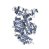
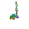
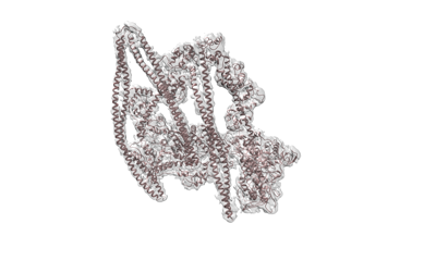
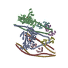
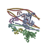
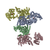
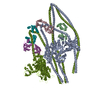
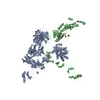
| Title | Structure of the autoinhibited state of smooth muscle myosin-2 | |||||||||
 Map data Map data | ||||||||||
 Sample Sample |
| |||||||||
 Keywords Keywords | Muscle contraction / ATPase / autoinhibition / 10S / CONTRACTILE PROTEIN | |||||||||
| Function / homology |  Function and homology information Function and homology informationRHO GTPases activate PAKs / myosin II filament / Smooth Muscle Contraction / elastic fiber assembly / skeletal muscle myosin thick filament assembly / myofibril assembly / myosin light chain binding / myosin II binding / smooth muscle contraction / muscle myosin complex ...RHO GTPases activate PAKs / myosin II filament / Smooth Muscle Contraction / elastic fiber assembly / skeletal muscle myosin thick filament assembly / myofibril assembly / myosin light chain binding / myosin II binding / smooth muscle contraction / muscle myosin complex / actomyosin / myosin filament / cardiac muscle cell development / actomyosin structure organization / myosin II complex / structural constituent of muscle / microfilament motor activity / myosin heavy chain binding / myofibril / stress fiber / ADP binding / actin filament binding / actin binding / calmodulin binding / calcium ion binding / magnesium ion binding / ATP binding / cytosol / cytoplasm Similarity search - Function | |||||||||
| Biological species |  | |||||||||
| Method | single particle reconstruction / cryo EM / Resolution: 3.4 Å | |||||||||
 Authors Authors | Heissler SM / Arora AS | |||||||||
 Citation Citation | Journal: Sci Adv / Year: 2021 Title: Cryo-EM structure of the autoinhibited state of myosin-2. Authors: Sarah M Heissler / Amandeep S Arora / Neil Billington / James R Sellers / Krishna Chinthalapudi /  Abstract: We solved the near-atomic resolution structure of smooth muscle myosin-2 in the autoinhibited state (10) using single-particle cryo–electron microscopy. The 3.4-Å structure reveals the precise ...We solved the near-atomic resolution structure of smooth muscle myosin-2 in the autoinhibited state (10) using single-particle cryo–electron microscopy. The 3.4-Å structure reveals the precise molecular architecture of 10 and the structural basis for myosin-2 regulation. We reveal the position of the phosphorylation sites that control myosin autoinhibition and activation by phosphorylation of the regulatory light chain. Further, we present a previously unidentified conformational state in myosin-2 that traps ADP and P produced by the hydrolysis of ATP in the active site. This noncanonical state represents a branch of the myosin enzyme cycle and explains the autoinhibition of the enzyme function of 10 along with its reduced affinity for actin. Together, our structure defines the molecular mechanisms that drive 10 formation, stabilization, and relief by phosphorylation of the regulatory light chain. | |||||||||
| History |
|
- Structure visualization
| Movie |
Movie viewer |
|---|---|
| Structure viewer | EM map: SurfViewMolmilJmol/JSmol |
| Supplemental images |
- Downloads & links
-EMDB archive
| Map data | emd_23810.map.gz | 251.8 MB | EMDB map data format | |
|---|---|---|---|---|
| Header (meta data) | emd-23810-v30.xmlemd-23810.xml | 16.6 KB 16.6 KB | Display Display | EMDB header |
| Images |  emd_23810.png emd_23810.png | 52.5 KB | ||
| Filedesc metadata | emd-23810.cif.gz | 7 KB | ||
| Archive directory |  http://ftp.pdbj.org/pub/emdb/structures/EMD-23810ftp://ftp.pdbj.org/pub/emdb/structures/EMD-23810 http://ftp.pdbj.org/pub/emdb/structures/EMD-23810ftp://ftp.pdbj.org/pub/emdb/structures/EMD-23810 | HTTPS FTP |
-Related structure data
| Related structure data |  7mf3MC M: atomic model generated by this map C: citing same article ( |
|---|---|
| Similar structure data |

-Links
| EMDB pages | EMDB (EBI/PDBe) / EMDataResource |
|---|---|
| Related items in Molecule of the Month |


-Map
| File | Download / File: emd_23810.map.gz / Format: CCP4 / Size: 512 MB / Type: IMAGE STORED AS FLOATING POINT NUMBER (4 BYTES) | ||||||||||||||||||||||||||||||||||||||||||||||||||||||||||||
|---|---|---|---|---|---|---|---|---|---|---|---|---|---|---|---|---|---|---|---|---|---|---|---|---|---|---|---|---|---|---|---|---|---|---|---|---|---|---|---|---|---|---|---|---|---|---|---|---|---|---|---|---|---|---|---|---|---|---|---|---|---|
| Projections & slices | Image control
Images are generated by Spider. | ||||||||||||||||||||||||||||||||||||||||||||||||||||||||||||
| Voxel size | X=Y=Z: 0.899 Å | ||||||||||||||||||||||||||||||||||||||||||||||||||||||||||||
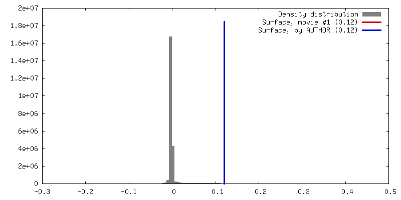
| Density |
| ||||||||||||||||||||||||||||||||||||||||||||||||||||||||||||
| Symmetry | Space group: 1 | ||||||||||||||||||||||||||||||||||||||||||||||||||||||||||||
| Details | EMDB XML:
CCP4 map header:
| ||||||||||||||||||||||||||||||||||||||||||||||||||||||||||||
 Z (Sec.)
 Y (Row.)
 X (Col.)


-Supplemental data
- Sample components
-Entire : Smooth muscle myosin-2 10S complex
| Entire | Name: Smooth muscle myosin-2 10S complex |
|---|---|
| Components |
|
-Supramolecule #1: Smooth muscle myosin-2 10S complex
| Supramolecule | Name: Smooth muscle myosin-2 10S complex / type: complex / ID: 1 / Parent: 0 / Macromolecule list: #1-#3 |
|---|---|
| Source (natural) | Organism: |
| Molecular weight | Theoretical: 531.256 KDa |
-Macromolecule #1: Myosin-11
| Macromolecule | Name: Myosin-11 / type: protein_or_peptide / ID: 1 / Number of copies: 4 / Enantiomer: LEVO |
|---|---|
| Source (natural) | Organism: |
| Molecular weight | Theoretical: 229.018953 KDa |
| Sequence | String: SQKPLSDDEK FLFVDKNFVN NPLAQADWSA KKLVWVPSEK HGFEAASIKE EKGDEVTVEL QENGKKVTLS KDDIQKMNPP KFSKVEDMA ELTCLNEASV LHNLRERYFS GLIYTYSGLF CVVINPYKQL PIYSEKIIDM YKGKKRHEMP PHIYAIADTA Y RSMLQDRE ...String: SQKPLSDDEK FLFVDKNFVN NPLAQADWSA KKLVWVPSEK HGFEAASIKE EKGDEVTVEL QENGKKVTLS KDDIQKMNPP KFSKVEDMA ELTCLNEASV LHNLRERYFS GLIYTYSGLF CVVINPYKQL PIYSEKIIDM YKGKKRHEMP PHIYAIADTA Y RSMLQDRE DQSILCTGES GAGKTENTKK VIQYLAVVAS SHKGKKDTSI TQGPSFSYGE LEKQLLQANP ILEAFGNAKT VK NDNSSRF GKFIRINFDV TGYIVGANIE TYLLEKSRAI RQAKDERTFH IFYYLIAGAS EQMRNDLLLE GFNNYTFLSN GHV PIPAQQ DDEMFQETLE AMTIMGFTEE EQTSILRVVS SVLQLGNIVF KKERNTDQAS MPDNTAAQKV CHLMGINVTD FTRS ILTPR IKVGRDVVQK AQTKEQADFA IEALAKAKFE RLFRWILTRV NKALDKTKRQ GASFLGILDI AGFEIFEINS FEQLC INYT NEKLQQLFNH TMFILEQEEY QREGIEWNFI DFGLDLQPCI ELIERPTNPP GVLALLDEEC WFPKATDTSF VEKLIQ EQG NHAKFQKSKQ LKDKTEFCIL HYAGKVTYNA SAWLTKNMDP LNDNVTSLLN QSSDKFVADL WKDVDRIVGL DQMAKMT ES SLPSASKTKK GMFRTVGQLY KEQLTKLMTT LRNTNPNFVR CIIPNHEKRA GKLDAHLVLE QLRCNGVLEG IRICRQGF P NRIVFQEFRQ RYEILAANAI PKGFMDGKQA CILMIKALEL DPNLYRIGQS KIFFRTGVLA HLEEERDLKI TDVIIAFQA QCRGYLARKA FAKRQQQLTA MKVIQRNCAA YLKLRNWQWW RLFTKVKPLL QVTRQEEEMQ AKDEELQRTK ERQQKAEAEL KELEQKHTQ LCEEKNLLQE KLQAETELYA EAEEMRVRLA AKKQELEEIL HEMEARIEEE EERSQQLQAE KKKMQQQMLD L EEQLEEEE AARQKLQLEK VTADGKIKKM EDDILIMEDQ NNKLTKERKL LEERVSDLTT NLAEEEEKAK NLTKLKNKHE SM ISELEVR LKKEEKSRQE LEKIKRKLEG ESSDLHEQIA ELQAQIAELK AQLAKKEEEL QAALARLEDE TSQKNNALKK IRE LESHIS DLQEDLESEK AARNKAEKQK RDLSEELEAL KTELEDTLDT TATQQELRAK REQEVTVLKR ALEEETRTHE AQVQ EMRQK HTQAVEELTE QLEQFKRAKA NLDKTKQTLE KDNADLANEI RSLSQAKQDV EHKKKKLEVQ LQDLQSKYSD GERVR TELN EKVHKLQIEV ENVTSLLNEA ESKNIKLTKD VATLGSQLQD TQELLQEETR QKLNVTTKLR QLEDDKNSLQ EQLDEE VEA KQNLERHIST LTIQLSDSKK KLQEFTATVE TMEEGKKKLQ REIESLTQQF EEKAASYDKL EKTKNRLQQE LDDLVVD LD NQRQLVSNLE KKQKKFDQML AEEKNISSKY ADERDRAEAE AREKETKALS LARALEEALE AKEELERTNK MLKAEMED L VSSKDDVGKN VHELEKSKRT LEQQVEEMKT QLEELEDELQ AAEDAKLRLE VNMQAMKSQF ERDLQARDEQ NEEKRRQLL KQLHEHETEL EDERKQRALA AAAKKKLEVD VKDLESQVDS ANKAREEAIK QLRKLQAQMK DYQRDLDDAR AAREEIFATA RENEKKAKN LEAELIQLQE DLAAAERARK QADLEKEEMA EELASANSGR TSLQDEKRRL EARIAQLEEE LDEEHSNIET M SDRMRKAV QQAEQLNNEL ATERATAQKN ENARQQLERQ NKELRSKLQE MEGAVKSKFK STIAALEAKI ASLEEQLEQE AR EKQAAAK TLRQKDKKLK DALLQVEDER KQAEQYKDQA EKGNLRLKQL KRQLEEAEEE SQRINANRRK LQRELDEATE SND ALGREV AALKSKLRRG NEPVSFAPPR RSGGRRVIEN ATDGGEEEID GRDGDFNGKA SE UniProtKB: Myosin-11 |
-Macromolecule #2: Myosin light polypeptide 6
| Macromolecule | Name: Myosin light polypeptide 6 / type: protein_or_peptide / ID: 2 / Number of copies: 2 / Enantiomer: LEVO |
|---|---|
| Source (natural) | Organism: |
| Molecular weight | Theoretical: 16.873025 KDa |
| Sequence | String: CDFSEEQTAE FKEAFQLFDR TGDGKILYSQ CGDVMRALGQ NPTNAEVMKV LGNPKSDEMN LKTLKFEQFL PMMQTIAKNK DQGCFEDYV EGLRVFDKEG NGTVMGAEIR HVLVTLGEKM TEEEVEQLVA GHEDSNGCIN YEELVRMVLS G UniProtKB: Myosin light polypeptide 6 |
-Macromolecule #3: Myosin regulatory light chain 2, smooth muscle major isoform
| Macromolecule | Name: Myosin regulatory light chain 2, smooth muscle major isoform type: protein_or_peptide / ID: 3 / Number of copies: 2 / Enantiomer: LEVO |
|---|---|
| Source (natural) | Organism: |
| Molecular weight | Theoretical: 19.741047 KDa |
| Sequence | String: SSKRAKAKTT KKRPQRATSN VFAMFDQSQI QEFKEAFNMI DQNRDGFIDK EDLHDMLASM GKNPTDEYLE GMMSEAPGPI NFTMFLTMF GEKLNGTDPE DVIRNAFACF DEEASGFIHE DHLRELLTTM GDRFTDEEVD EMYREAPIDK KGNFNYVEFT R ILKHGAKD KDD UniProtKB: Myosin regulatory light chain 2, smooth muscle major isoform |
-Macromolecule #4: ADENOSINE-5'-DIPHOSPHATE
| Macromolecule | Name: ADENOSINE-5'-DIPHOSPHATE / type: ligand / ID: 4 / Number of copies: 2 / Formula: ADP |
|---|---|
| Molecular weight | Theoretical: 427.201 Da |
| Chemical component information |  ChemComp-ADP: |
-Macromolecule #5: PHOSPHATE ION
| Macromolecule | Name: PHOSPHATE ION / type: ligand / ID: 5 / Number of copies: 2 / Formula: PO4 |
|---|---|
| Molecular weight | Theoretical: 94.971 Da |
| Chemical component information |  ChemComp-PO4: |
-Macromolecule #6: MAGNESIUM ION
| Macromolecule | Name: MAGNESIUM ION / type: ligand / ID: 6 / Number of copies: 4 / Formula: MG |
|---|---|
| Molecular weight | Theoretical: 24.305 Da |
-Experimental details
-Structure determination
| Method | cryo EM |
|---|---|
 Processing Processing | single particle reconstruction |
| Aggregation state | tissue |
-Sample preparation
| Buffer | pH: 7.3 / Component - Concentration: 150.0 mM / Component - Formula: NaCl / Component - Name: sodium chloride / Details: 150 mM NaCl, 1mM EGTA, 2mM MgCl2 |
|---|---|
| Grid | Model: C-flat-1.2/1.3 / Material: GOLD / Mesh: 300 / Support film - Material: CARBON / Support film - topology: HOLEY / Pretreatment - Type: GLOW DISCHARGE / Pretreatment - Time: 45 sec. / Pretreatment - Atmosphere: AIR |
| Vitrification | Cryogen name: ETHANE / Chamber humidity: 95 % / Chamber temperature: 292 K / Instrument: LEICA EM GP |
| Details | 1 mg/ml |
- Electron microscopy
| Microscope | FEI TITAN KRIOS |
|---|---|
| Specialist optics | Energy filter - Name: GIF Bioquantum / Energy filter - Slit width: 20 eV |
| Image recording | Film or detector model: GATAN K3 BIOQUANTUM (6k x 4k) / Number grids imaged: 1 / Average electron dose: 65.0 e/Å2 |
| Electron beam | Acceleration voltage: 300 kV / Electron source:  FIELD EMISSION GUN FIELD EMISSION GUN |
| Electron optics | Illumination mode: OTHER / Imaging mode: BRIGHT FIELD / Cs: 0.01 mm / Nominal magnification: 81000 |
| Sample stage | Specimen holder model: FEI TITAN KRIOS AUTOGRID HOLDER |
| Experimental equipment |  Model: Titan Krios / Image courtesy: FEI Company |
-Image processing
| Startup model | Type of model: PDB ENTRY PDB model - PDB ID: |
|---|---|
| Final reconstruction | Number classes used: 1 / Applied symmetry - Point group: C1 (asymmetric) / Resolution.type: BY AUTHOR / Resolution: 3.4 Å / Resolution method: FSC 0.143 CUT-OFF / Software - Name: cryoSPARC / Number images used: 234464 |
| Initial angle assignment | Type: OTHER / Software - Name: cryoSPARC |
| Final angle assignment | Type: OTHER / Software - Name: cryoSPARC |