 Movie
Movie Controller

[English] 日本語
 Yorodumi
- PDB-3cw2: Crystal structure of the intact archaeal translation initiation f... -
+ Open data

- Basic information
| Entry | Database: PDB / ID: 3cw2 | ||||||
|---|---|---|---|---|---|---|---|
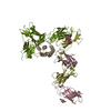
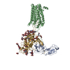
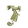
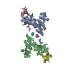
| Title | Crystal structure of the intact archaeal translation initiation factor 2 from Sulfolobus solfataricus . | ||||||
 Components Components |
| ||||||
 Keywords Keywords | TRANSLATION / AIF2 / INTACT AIF2 / INITIATION FACTOR 2 ALPHA SUBUNIT / INITIATION FACTOR 2 BETA SUBUNIT / INITIATION FACTOR 2 GAMMA SUBUNIT / INITIATION OF THE TRANSLATION / Initiation factor / Protein biosynthesis / RNA-binding / GTP-binding / Nucleotide-binding | ||||||
| Function / homology |  Function and homology information Function and homology informationformation of translation preinitiation complex / protein-synthesizing GTPase / translation elongation factor activity / translation initiation factor activity / translational initiation / ribosome binding / tRNA binding / GTPase activity / GTP binding / RNA binding / metal ion binding Similarity search - Function | ||||||
| Biological species |   Sulfolobus solfataricus (archaea) Sulfolobus solfataricus (archaea) | ||||||
| Method |  X-RAY DIFFRACTION / SYNCHROTRON / MOLECULAR REPLACEMENT / Resolution: 2.8 Å X-RAY DIFFRACTION / SYNCHROTRON / MOLECULAR REPLACEMENT / Resolution: 2.8 Å | ||||||
 Authors Authors | Stolboushkina, E.A. / Nikonov, S.V. / Nikulin, A.D. / Blaesi, U. / Manstein, D.J. / Fedorov, R.V. / Garber, M.B. / Nikonov, O.S. | ||||||
 Citation Citation | Journal: J.Mol.Biol. / Year: 2008 Title: Crystal structure of the intact archaeal translation initiation factor 2 demonstrates very high conformational flexibility in the alpha- and beta-subunits. Authors: Stolboushkina, E. / Nikonov, S. / Nikulin, A. / Blasi, U. / Manstein, D.J. / Fedorov, R. / Garber, M. / Nikonov, O. | ||||||
| History |
|
- Structure visualization
| Structure viewer | Molecule: MolmilJmol/JSmol |
|---|
- Downloads & links
-Download
| PDBx/mmCIF format | 3cw2.cif.gz | 587.2 KB | Display | PDBx/mmCIF format |
|---|---|---|---|---|
| PDB format | pdb3cw2.ent.gz | 481.4 KB | Display | PDB format |
| PDBx/mmJSON format | 3cw2.json.gz | Tree view | PDBx/mmJSON format | |
| Others |  Other downloads Other downloads |
-Validation report
| Arichive directory | https://data.pdbj.org/pub/pdb/validation_reports/cw/3cw2ftp://data.pdbj.org/pub/pdb/validation_reports/cw/3cw2 | HTTPS FTP |
|---|
-Related structure data
| Related structure data |  2plfS S: Starting model for refinement |
|---|---|
| Similar structure data |


-Links
 PDBj
PDBj


- Assembly
| Deposited unit | 
| ||||||||
|---|---|---|---|---|---|---|---|---|---|
| 1 | 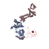
| ||||||||
| 2 | 
| ||||||||
| 3 | 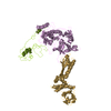
| ||||||||
| 4 | 
| ||||||||
| Unit cell |
|
-Components
| #1: Protein | Mass: 45849.230 Da / Num. of mol.: 4 / Fragment: aIF2gamma subunit Source method: isolated from a genetically manipulated source Source: (gene. exp.) Sulfolobus solfataricus (archaea) / Gene: eif2g, SSO0412 / Plasmid: pET11d / Production host:  #2: Protein | Mass: 30432.355 Da / Num. of mol.: 4 / Fragment: aIF2alpha subunit Source method: isolated from a genetically manipulated source Source: (gene. exp.) Sulfolobus solfataricus (archaea) / Gene: eif2a, SSO1050 / Plasmid: pET11c / Production host: #3: Protein | Mass: 15942.740 Da / Num. of mol.: 4 / Fragment: aIF2beta subunit Source method: isolated from a genetically manipulated source Source: (gene. exp.) Sulfolobus solfataricus (archaea) / Gene: eif2b, SSO2381 / Plasmid: pET11d / Production host: Has protein modification | Y | |
|---|
-Experimental details
-Experiment
| Experiment | Method: X-RAY DIFFRACTION / Number of used crystals: 1 |
|---|
- Sample preparation
| Crystal | Density Matthews: 2.82 Å3/Da / Density % sol: 56.39 % |
|---|---|
| Crystal grow | Temperature: 295 K / Method: vapor diffusion, hanging drop / pH: 8.5 Details: 0.1M Tris HCl, 0.72M sodium formate, 9% PEG 8000, 9% PEG 1000, 1% monomethyl ether polyethylene glycol 5000 , pH 8.5, VAPOR DIFFUSION, HANGING DROP, temperature 295K |
-Data collection
| Diffraction | Mean temperature: 100 K |
|---|---|
| Diffraction source | Source: SYNCHROTRON / Site: EMBL/DESY, HAMBURG  / Beamline: X12 / Wavelength: 0.8423 Å / Beamline: X12 / Wavelength: 0.8423 Å |
| Detector | Type: MAR scanner 345 mm plate / Detector: IMAGE PLATE / Date: Feb 16, 2007 |
| Radiation | Monochromator: MIRRORS / Protocol: SINGLE WAVELENGTH / Monochromatic (M) / Laue (L): M / Scattering type: x-ray |
| Radiation wavelength | Wavelength: 0.8423 Å / Relative weight: 1 |
| Reflection twin | Operator: h,-k,-l / Fraction: 0.459 |
| Reflection | Resolution: 2.8→19.913 Å / Num. all: 98811 / Num. obs: 95812 / % possible obs: 95.3 % / Observed criterion σ(F): 0 / Observed criterion σ(I): 0 / Redundancy: 3.16 % / Rmerge(I) obs: 0.069 / Net I/σ(I): 10.71 |
| Reflection shell | Resolution: 2.8→2.85 Å / % possible all: 90.9 |
- Processing
| Software |
| |||||||||||||||||||||||||
|---|---|---|---|---|---|---|---|---|---|---|---|---|---|---|---|---|---|---|---|---|---|---|---|---|---|---|
| Refinement | Method to determine structure: MOLECULAR REPLACEMENT Starting model: PDB ENTRY 2PLF Resolution: 2.8→19.9118 Å / Cross valid method: THROUGHOUT / σ(F): 0 / Stereochemistry target values: Engh & Huber
| |||||||||||||||||||||||||
| Displacement parameters | Biso mean: 71.899 Å2 | |||||||||||||||||||||||||
| Refinement step | Cycle: LAST / Resolution: 2.8→19.9118 Å
|
