 Movie
Movie Controller

[English] 日本語
 Yorodumi
- PDB-2plf: The structure of aIF2gamma subunit from the archaeon Sulfolobus s... -
+ Open data

- Basic information
| Entry | Database: PDB / ID: 2plf | ||||||
|---|---|---|---|---|---|---|---|
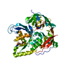
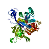
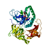
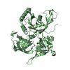
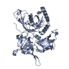
| Title | The structure of aIF2gamma subunit from the archaeon Sulfolobus solfataricus in the nucleotide-free form. | ||||||
 Components Components | Translation initiation factor 2 gamma subunit | ||||||
 Keywords Keywords | TRANSLATION / aIF2 / Initiation factor 2 gamma subunit / Initiation of the translation / nucleotide binding | ||||||
| Function / homology |  Function and homology information Function and homology informationformation of translation preinitiation complex / protein-synthesizing GTPase / translation elongation factor activity / translation initiation factor activity / tRNA binding / GTPase activity / GTP binding / metal ion binding Similarity search - Function | ||||||
| Biological species |   Sulfolobus solfataricus (archaea) Sulfolobus solfataricus (archaea) | ||||||
| Method |  X-RAY DIFFRACTION / SYNCHROTRON / MOLECULAR REPLACEMENT / Resolution: 2.9 Å X-RAY DIFFRACTION / SYNCHROTRON / MOLECULAR REPLACEMENT / Resolution: 2.9 Å | ||||||
 Authors Authors | Nikonov, O.S. / Stolboushkina, E.A. / Nikulin, A.D. / Hasenohrl, D. / Blaesi, U. / Manstein, D.J. / Fedorov, R.V. / Garber, M.B. / Nikonov, S.V. | ||||||
 Citation Citation | Journal: J.Mol.Biol. / Year: 2007 Title: New Insights into the Interactions of the Translation Initiation Factor 2 from Archaea with Guanine Nucleotides and Initiator tRNA. Authors: Nikonov, O. / Stolboushkina, E. / Nikulin, A. / Hasenohrl, D. / Blasi, U. / Manstein, D.J. / Fedorov, R. / Garber, M. / Nikonov, S. | ||||||
| History |
|
- Structure visualization
| Structure viewer | Molecule: MolmilJmol/JSmol |
|---|
- Downloads & links
-Download
| PDBx/mmCIF format | 2plf.cif.gz | 93.9 KB | Display | PDBx/mmCIF format |
|---|---|---|---|---|
| PDB format | pdb2plf.ent.gz | 71.4 KB | Display | PDB format |
| PDBx/mmJSON format | 2plf.json.gz | Tree view | PDBx/mmJSON format | |
| Others |  Other downloads Other downloads |
-Validation report
| Arichive directory | https://data.pdbj.org/pub/pdb/validation_reports/pl/2plfftp://data.pdbj.org/pub/pdb/validation_reports/pl/2plf | HTTPS FTP |
|---|
-Related structure data
| Related structure data |  2pmdC  2ahoS S: Starting model for refinement C: citing same article ( |
|---|---|
| Similar structure data |


-Links
 PDBj
PDBj


- Assembly
| Deposited unit | 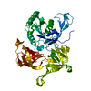
| ||||||||
|---|---|---|---|---|---|---|---|---|---|
| 1 |
| ||||||||
| Unit cell |
|
-Components
| #1: Protein | Mass: 45718.035 Da / Num. of mol.: 1 Source method: isolated from a genetically manipulated source Source: (gene. exp.) Sulfolobus solfataricus (archaea) / Gene: eif2g / Plasmid: pET11d / Production host:  |
|---|---|
| #2: Water | ChemComp-HOH /  Mass: 18.015 Da / Num. of mol.: 98 / Source method: isolated from a natural source / Formula: H2O Mass: 18.015 Da / Num. of mol.: 98 / Source method: isolated from a natural source / Formula: H2O |
| Has protein modification | Y |
-Experimental details
-Experiment
| Experiment | Method: X-RAY DIFFRACTION / Number of used crystals: 2 |
|---|
- Sample preparation
| Crystal | Density Matthews: 4.72 Å3/Da / Density % sol: 73.96 % |
|---|---|
| Crystal grow | Temperature: 295 K / pH: 4.5 Details: natrium malonate dihydrate, glycine,CdCl2., pH 4.5, VAPOR DIFFUSION, HANGING DROP, temperature 295K, pH 4.50 |
-Data collection
| Diffraction | Mean temperature: 100 K |
|---|---|
| Diffraction source | Source: SYNCHROTRON / Site: EMBL/DESY, HAMBURG  / Beamline: X12 / Wavelength: 1 / Beamline: X12 / Wavelength: 1 |
| Detector | Type: MARMOSAIC 225 mm CCD / Detector: CCD / Date: Jul 3, 2006 |
| Radiation | Protocol: SINGLE WAVELENGTH / Monochromatic (M) / Laue (L): M / Scattering type: x-ray |
| Radiation wavelength | Wavelength: 1 Å / Relative weight: 1 |
| Reflection | Resolution: 2.9→20 Å / Num. obs: 19573 / % possible obs: 98.7 % / Observed criterion σ(I): 0 / Redundancy: 11.9 % / Rmerge(I) obs: 0.105 |
| Reflection shell | Resolution: 2.9→3 Å / % possible all: 97 |
- Processing
| Software |
| |||||||||||||||||||||||||
|---|---|---|---|---|---|---|---|---|---|---|---|---|---|---|---|---|---|---|---|---|---|---|---|---|---|---|
| Refinement | Method to determine structure: MOLECULAR REPLACEMENT Starting model: GAMMA SUBUNIT FORM THE AIF2 ALPHA-GAMMA DIMER. PDB ENTRY: 2AHO Resolution: 2.9→20 Å / Isotropic thermal model: ISOTROPIC / Cross valid method: THROUGHOUT / σ(F): 2 / Stereochemistry target values: ENGH & HUBER
| |||||||||||||||||||||||||
| Displacement parameters | Biso mean: 58.85 Å2 | |||||||||||||||||||||||||
| Refinement step | Cycle: LAST / Resolution: 2.9→20 Å
|
