 Movie
Movie Controller

[English] 日本語
 Yorodumi
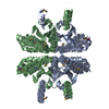
- PDB-2a8d: Haemophilus influenzae beta-carbonic anhydrase complexed with bic... -
+ Open data

- Basic information
| Entry | Database: PDB / ID: 2a8d | ||||||
|---|---|---|---|---|---|---|---|
| Title | Haemophilus influenzae beta-carbonic anhydrase complexed with bicarbonate | ||||||
 Components Components | Carbonic anhydrase 2 | ||||||
 Keywords Keywords | LYASE / x-ray structure / carbonic anhydrase / protein-bicarbonate complex | ||||||
| Function / homology |  Function and homology information Function and homology informationcarbon utilization / carbonic anhydrase / carbonate dehydratase activity / zinc ion binding Similarity search - Function | ||||||
| Biological species |  Haemophilus influenzae (bacteria) Haemophilus influenzae (bacteria) | ||||||
| Method |  X-RAY DIFFRACTION / MOLECULAR REPLACEMENT / Resolution: 2.2 Å X-RAY DIFFRACTION / MOLECULAR REPLACEMENT / Resolution: 2.2 Å | ||||||
 Authors Authors | Cronk, J.D. / Rowlett, R.S. / Zhang, K.Y.J. / Tu, C. / Endrizzi, J.A. / Lee, J. / Gareiss, P.C. / Preiss, J.R. | ||||||
 Citation Citation | Journal: Biochemistry / Year: 2006 Title: Identification of a Novel Noncatalytic Bicarbonate Binding Site in Eubacterial beta-Carbonic Anhydrase Authors: Cronk, J.D. / Rowlett, R.S. / Zhang, K.Y.J. / Tu, C. / Endrizzi, J.A. / Lee, J. / Gareiss, P.C. / Preiss, J.R. | ||||||
| History |
|
- Structure visualization
| Structure viewer | Molecule: MolmilJmol/JSmol |
|---|
- Downloads & links
-Download
| PDBx/mmCIF format | 2a8d.cif.gz | 277.4 KB | Display | PDBx/mmCIF format |
|---|---|---|---|---|
| PDB format | pdb2a8d.ent.gz | 224.8 KB | Display | PDB format |
| PDBx/mmJSON format | 2a8d.json.gz | Tree view | PDBx/mmJSON format | |
| Others |  Other downloads Other downloads |
-Validation report
| Arichive directory | https://data.pdbj.org/pub/pdb/validation_reports/a8/2a8dftp://data.pdbj.org/pub/pdb/validation_reports/a8/2a8d | HTTPS FTP |
|---|
-Related structure data
| Related structure data | 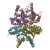 2a8cC 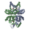 2esfC  1i6pS C: citing same article ( S: Starting model for refinement |
|---|---|
| Similar structure data |


-Links
 PDBj
PDBj


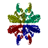
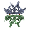
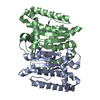
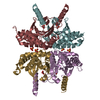
- Assembly
| Deposited unit | 
| ||||||||||||||||||
|---|---|---|---|---|---|---|---|---|---|---|---|---|---|---|---|---|---|---|---|
| 1 | 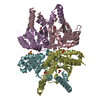
| ||||||||||||||||||
| 2 | 
| ||||||||||||||||||
| Unit cell |
| ||||||||||||||||||
| Components on special symmetry positions |
| ||||||||||||||||||
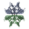
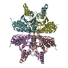
| Details | Chains C, D, E, and F comprise a biological assembly. A second biological assembly can be formed from chains A and B by the operation -x, y, -z |
-Components
| #1: Protein | Mass: 26286.984 Da / Num. of mol.: 6 Source method: isolated from a genetically manipulated source Source: (gene. exp.) Haemophilus influenzae (bacteria) / Gene: can / Plasmid: pTrc99a / Production host: #2: Chemical | ChemComp-ZN /   Mass: 65.409 Da / Num. of mol.: 6 / Source method: obtained synthetically / Formula: Zn Mass: 65.409 Da / Num. of mol.: 6 / Source method: obtained synthetically / Formula: Zn#3: Chemical | ChemComp-BCT /   Mass: 61.017 Da / Num. of mol.: 6 / Source method: obtained synthetically / Formula: CHO3 / Comment: pH buffer*YM Mass: 61.017 Da / Num. of mol.: 6 / Source method: obtained synthetically / Formula: CHO3 / Comment: pH buffer*YM#4: Chemical | ChemComp-SO4 /   Mass: 96.063 Da / Num. of mol.: 6 / Source method: obtained synthetically / Formula: SO4 Mass: 96.063 Da / Num. of mol.: 6 / Source method: obtained synthetically / Formula: SO4#5: Water | ChemComp-HOH / |  Mass: 18.015 Da / Num. of mol.: 318 / Source method: isolated from a natural source / Formula: H2O Mass: 18.015 Da / Num. of mol.: 318 / Source method: isolated from a natural source / Formula: H2O |
|---|
-Experimental details
-Experiment
| Experiment | Method: X-RAY DIFFRACTION / Number of used crystals: 1 |
|---|
- Sample preparation
| Crystal | Density Matthews: 2.78 Å3/Da / Density % sol: 55.7 % |
|---|---|
| Crystal grow | Temperature: 277 K / Method: vapor diffusion, hanging drop / pH: 7.5 Details: ammonium sulfate, PEG 400, HEPES, pH 7.5, VAPOR DIFFUSION, HANGING DROP, temperature 277K |
-Data collection
| Diffraction | Mean temperature: 95 K |
|---|---|
| Diffraction source | Source: ROTATING ANODE / Type: RIGAKU RU200 / Wavelength: 1.5418 Å |
| Detector | Type: RIGAKU RAXIS IV / Detector: IMAGE PLATE / Date: Dec 17, 2002 |
| Radiation | Monochromator: yale mirrors / Protocol: SINGLE WAVELENGTH / Monochromatic (M) / Laue (L): M / Scattering type: x-ray |
| Radiation wavelength | Wavelength: 1.5418 Å / Relative weight: 1 |
| Reflection | Resolution: 2.2→30 Å / Num. all: 86683 / Num. obs: 86531 / % possible obs: 99.6 % / Observed criterion σ(I): 5 / Redundancy: 2.87 % / Biso Wilson estimate: 40.77 Å2 / Rsym value: 0.043 / Net I/σ(I): 22 |
| Reflection shell | Resolution: 2.2→2.28 Å / Redundancy: 2.29 % / Mean I/σ(I) obs: 3.18 / Num. unique all: 8444 / Rsym value: 0.262 / % possible all: 97.4 |
- Processing
| Software |
| ||||||||||||||||||||||||||||||||||||
|---|---|---|---|---|---|---|---|---|---|---|---|---|---|---|---|---|---|---|---|---|---|---|---|---|---|---|---|---|---|---|---|---|---|---|---|---|---|
| Refinement | Method to determine structure: MOLECULAR REPLACEMENT Starting model: PDB entry 1I6P Resolution: 2.2→30 Å / Data cutoff high absF: 10000 / Data cutoff low absF: 0 / Isotropic thermal model: anisotropic / Cross valid method: THROUGHOUT / σ(F): 0 / Stereochemistry target values: Engh & Huber
| ||||||||||||||||||||||||||||||||||||
| Displacement parameters |
| ||||||||||||||||||||||||||||||||||||
| Refine analyze |
| ||||||||||||||||||||||||||||||||||||
| Refinement step | Cycle: LAST / Resolution: 2.2→30 Å
| ||||||||||||||||||||||||||||||||||||
| Refine LS restraints |
| ||||||||||||||||||||||||||||||||||||
| LS refinement shell | Resolution: 2.2→2.28 Å
|
