 Movie
Movie Controller


+ Open data

- Basic information
| Entry | Database: PDB / ID: 1i6p | ||||||
|---|---|---|---|---|---|---|---|
| Title | CRYSTAL STRUCTURE OF E. COLI BETA CARBONIC ANHYDRASE (ECCA) | ||||||
 Components Components | CARBONIC ANHYDRASE | ||||||
 Keywords Keywords | LYASE / carbonic anhydrase / metalloenzyme / zinc coordination / pH-dependent activity / MAD phasing | ||||||
| Function / homology |  Function and homology information Function and homology informationcarbon utilization / carbonic anhydrase / carbonate dehydratase activity / protein homotetramerization / zinc ion binding / identical protein binding / cytosol Similarity search - Function | ||||||
| Biological species |  | ||||||
| Method |  X-RAY DIFFRACTION / MOLECULAR REPLACEMENT / Resolution: 2 Å X-RAY DIFFRACTION / MOLECULAR REPLACEMENT / Resolution: 2 Å | ||||||
 Authors Authors | Cronk, J.D. / Endrizzi, J.A. / Cronk, M.R. / O'Neill, J.W. / Zhang, K.Y.J. | ||||||
 Citation Citation | Journal: Protein Sci. / Year: 2001 Title: Crystal structure of E. coli beta-carbonic anhydrase, an enzyme with an unusual pH-dependent activity. Authors: Cronk, J.D. / Endrizzi, J.A. / Cronk, M.R. / O'neill, J.W. / Zhang, K.Y. #1: Journal: Acta Crystallogr.,Sect.D / Year: 2000Title: Cloning, crystallization and preliminary characterization of a beta carbonic anhydrase from Escherichia coli Authors: Cronk, J.D. / O'Neill, J.W. / Cronk, M.R. / Endrizzi, J.A. / Zhang, K.Y.J. | ||||||
| History |
| ||||||
| Remark 99 | DENSITY FOR THE FOLLOWING ATOMS WITHIN THE MAIN CHAIN IS RELATIVELY WEAK AND LIKELY REFLECTS ...DENSITY FOR THE FOLLOWING ATOMS WITHIN THE MAIN CHAIN IS RELATIVELY WEAK AND LIKELY REFLECTS INCREASED LOCAL CONFORMATIONAL FLEXIBILITY OF THE MAIN CHAIN IN THIS REGION. GLN A 31 CA ALA A 32 N ALA A 32 CA DENSITY FOR THE SIDE CHAINS OF THE LISTED RESIDUES IS RELATIVELY POORLY DEFINED. THE MODEL REPRESENTS THE MOST PROBABLE CONFORMATIONS FOR THESE RESIDUES BASED ON THE OBSERVED DENSITY. GLU A 20 GLU A 21 LYS A 28 LEU A 29 GLN A 31 ALA A 32 GLN A 33 LYS A 34 GLU A 112 GLU A 140 ARG A 141 LYS A 169 LYS A 173 ARG A 189 ARG A 198 GLU A 199 LYS A 213 LEU A 214 LYS A 215 |
- Structure visualization
| Structure viewer | Molecule: MolmilJmol/JSmol |
|---|
- Downloads & links
-Download
| PDBx/mmCIF format | 1i6p.cif.gz | 57.7 KB | Display | PDBx/mmCIF format |
|---|---|---|---|---|
| PDB format | pdb1i6p.ent.gz | 41.2 KB | Display | PDB format |
| PDBx/mmJSON format | 1i6p.json.gz | Tree view | PDBx/mmJSON format | |
| Others |  Other downloads Other downloads |
-Validation report
| Arichive directory | https://data.pdbj.org/pub/pdb/validation_reports/i6/1i6pftp://data.pdbj.org/pub/pdb/validation_reports/i6/1i6p | HTTPS FTP |
|---|
-Related structure data
| Related structure data | 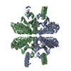 1i6oSC S: Starting model for refinement C: citing same article ( |
|---|---|
| Similar structure data |


-Links
 PDBj
PDBj
- Assembly
| Deposited unit | 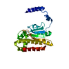
| ||||||||
|---|---|---|---|---|---|---|---|---|---|
| 1 | 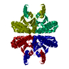
| ||||||||
| Unit cell |
| ||||||||
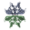
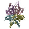
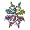
| Details | A tetramer constructed from the operations -y, -x, -z; y, x, -z; -1-x, 1-y, z is thought to exist in solution under physiological conditions. |
-Components
| #1: Protein | Mass: 25130.779 Da / Num. of mol.: 1 Source method: isolated from a genetically manipulated source Source: (gene. exp.) |
|---|---|
| #2: Chemical | ChemComp-ZN /   Mass: 65.409 Da / Num. of mol.: 1 / Source method: obtained synthetically / Formula: Zn Mass: 65.409 Da / Num. of mol.: 1 / Source method: obtained synthetically / Formula: Zn |
| #3: Water | ChemComp-HOH /  Mass: 18.015 Da / Num. of mol.: 62 / Source method: isolated from a natural source / Formula: H2O Mass: 18.015 Da / Num. of mol.: 62 / Source method: isolated from a natural source / Formula: H2O |
-Experimental details
-Experiment
| Experiment | Method: X-RAY DIFFRACTION / Number of used crystals: 1 |
|---|
- Sample preparation
| Crystal | Density Matthews: 2.009 Å3/Da / Density % sol: 36.4 % | ||||||||||||||||||||||||||||||||||||
|---|---|---|---|---|---|---|---|---|---|---|---|---|---|---|---|---|---|---|---|---|---|---|---|---|---|---|---|---|---|---|---|---|---|---|---|---|---|
| Crystal grow | Temperature: 293 K / Method: vapor diffusion, hanging drop / pH: 6.3 Details: ammonium sulfate, MES pH 6.3, VAPOR DIFFUSION, HANGING DROP, temperature 293K | ||||||||||||||||||||||||||||||||||||
| Crystal grow | *PLUS Temperature: 20 ℃ / pH: 8.3 | ||||||||||||||||||||||||||||||||||||
| Components of the solutions | *PLUS
|
-Data collection
| Diffraction | Mean temperature: 291 K |
|---|---|
| Diffraction source | Source: ROTATING ANODE / Type: RIGAKU RU200 / Wavelength: 1.5418 Å |
| Detector | Type: RIGAKU RAXIS IV / Detector: IMAGE PLATE / Date: Apr 8, 1999 / Details: mirrors |
| Radiation | Monochromator: mirrors / Protocol: SINGLE WAVELENGTH / Monochromatic (M) / Laue (L): M / Scattering type: x-ray |
| Radiation wavelength | Wavelength: 1.5418 Å / Relative weight: 1 |
| Reflection | Resolution: 2→40 Å / Num. all: 90500 / Num. obs: 13974 / % possible obs: 96.7 % / Observed criterion σ(F): 0 / Observed criterion σ(I): 0 / Redundancy: 6.5 % / Biso Wilson estimate: 15.6 Å2 / Rmerge(I) obs: 0.065 / Net I/σ(I): 20.4 |
| Reflection shell | Resolution: 2→2.03 Å / Redundancy: 3.5 % / Rmerge(I) obs: 0.309 / Mean I/σ(I) obs: 4.4 / % possible all: 96.9 |
- Processing
| Software |
| |||||||||||||||||||||||||
|---|---|---|---|---|---|---|---|---|---|---|---|---|---|---|---|---|---|---|---|---|---|---|---|---|---|---|
| Refinement | Method to determine structure: MOLECULAR REPLACEMENT Starting model: PDB ENTRY 1I6O Resolution: 2→42.21 Å / Rfactor Rfree error: 0.005 / Data cutoff high absF: 334555.69 / Data cutoff low absF: 0 / Isotropic thermal model: RESTRAINED / Cross valid method: THROUGHOUT / σ(F): 0 / σ(I): 0 / Stereochemistry target values: Engh & Huber
| |||||||||||||||||||||||||
| Solvent computation | Solvent model: FLAT MODEL / Bsol: 44.43 Å2 / ksol: 0.338 e/Å3 | |||||||||||||||||||||||||
| Displacement parameters | Biso mean: 28.8 Å2
| |||||||||||||||||||||||||
| Refine analyze |
| |||||||||||||||||||||||||
| Refinement step | Cycle: LAST / Resolution: 2→42.21 Å
| |||||||||||||||||||||||||
| Refine LS restraints |
| |||||||||||||||||||||||||
| LS refinement shell | Resolution: 2→2.13 Å / Rfactor Rfree error: 0.014 / Total num. of bins used: 6
| |||||||||||||||||||||||||
| Xplor file |
| |||||||||||||||||||||||||
| Software | *PLUS Name: CNS / Version: 1 / Classification: refinement | |||||||||||||||||||||||||
| Refinement | *PLUS σ(F): 0 / % reflection Rfree: 10.1 % | |||||||||||||||||||||||||
| Solvent computation | *PLUS | |||||||||||||||||||||||||
| Displacement parameters | *PLUS Biso mean: 28.8 Å2 | |||||||||||||||||||||||||
| Refine LS restraints | *PLUS
| |||||||||||||||||||||||||
| LS refinement shell | *PLUS Rfactor Rfree: 0.215 / % reflection Rfree: 10.2 % / Rfactor Rwork: 0.191 |
