National Institutes of Health/National Institute of Neurological Disorders and Stroke (NIH/NINDS)
R01NS088367
United States
National Institutes of Health/National Institute Of Allergy and Infectious Diseases (NIH/NIAID)
R01AI107121
United States
National Institutes of Health/National Institute of Neurological Disorders and Stroke (NIH/NINDS)
R01NS092662
United States
National Institutes of Health/National Institute of Neurological Disorders and Stroke (NIH/NINDS)
F31NS083336
United States
National Institutes of Health/National Institute of Neurological Disorders and Stroke (NIH/NINDS)
F32NS106730
United States
National Institutes of Health/National Cancer Institute (NIH/NCI)
T32CA060395
United States
Citation
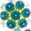
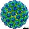
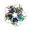
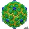
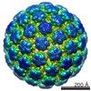
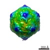
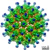
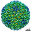
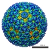
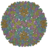
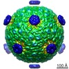
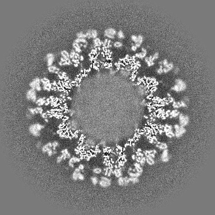
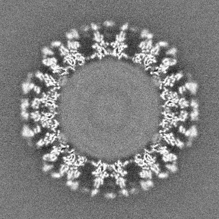
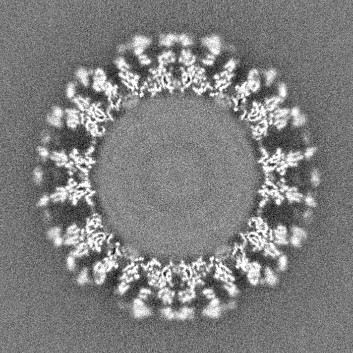
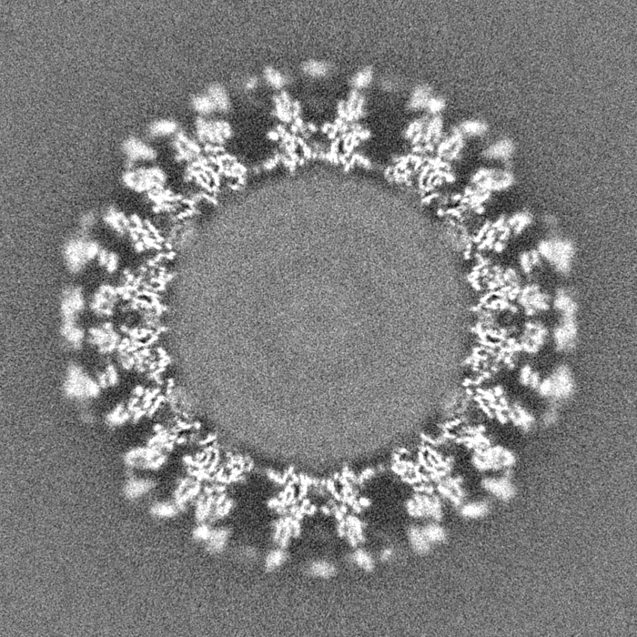
Journal: Elife / Year: 2020 Title: Antibody escape by polyomavirus capsid mutation facilitates neurovirulence. Authors: Matthew D Lauver / Daniel J Goetschius / Colleen S Netherby-Winslow / Katelyn N Ayers / Ge Jin / Daniel G Haas / Elizabeth L Frost / Sung Hyun Cho / Carol M Bator / Stephanie M Bywaters / ...Authors: Matthew D Lauver / Daniel J Goetschius / Colleen S Netherby-Winslow / Katelyn N Ayers / Ge Jin / Daniel G Haas / Elizabeth L Frost / Sung Hyun Cho / Carol M Bator / Stephanie M Bywaters / Neil D Christensen / Susan L Hafenstein / Aron E Lukacher / Abstract: JCPyV polyomavirus, a member of the human virome, causes progressive multifocal leukoencephalopathy (PML), an oft-fatal demyelinating brain disease in individuals receiving immunomodulatory therapies. ...JCPyV polyomavirus, a member of the human virome, causes progressive multifocal leukoencephalopathy (PML), an oft-fatal demyelinating brain disease in individuals receiving immunomodulatory therapies. Mutations in the major viral capsid protein, VP1, are common in JCPyV from PML patients (JCPyV-PML) but whether they confer neurovirulence or escape from virus-neutralizing antibody (nAb) in vivo is unknown. A mouse polyomavirus (MuPyV) with a sequence-equivalent JCPyV-PML VP1 mutation replicated poorly in the kidney, a major reservoir for JCPyV persistence, but retained the CNS infectivity, cell tropism, and neuropathology of the parental virus. This mutation rendered MuPyV resistant to a monoclonal Ab (mAb), whose specificity overlapped the endogenous anti-VP1 response. Using cryo-EM and a custom sub-particle refinement approach, we resolved an MuPyV:Fab complex map to 3.2 Å resolution. The structure revealed the mechanism of mAb evasion. Our findings demonstrate convergence between nAb evasion and CNS neurovirulence in vivo by a frequent JCPyV-PML VP1 mutation.
History
Deposition
Sep 9, 2020
-
Header (metadata) release
Sep 23, 2020
-
Map release
Sep 23, 2020
-
Update
Oct 28, 2020
-
Current status
Oct 28, 2020
Processing site: RCSB / Status: Released
-
Structure visualization
Movie
Surface view with section colored by density value
In the structure databanks used in Yorodumi, some data are registered as the other names, "COVID-19 virus" and "2019-nCoV". Here are the details of the virus and the list of structure data.
Jan 31, 2019. EMDB accession codes are about to change! (news from PDBe EMDB page)
EMDB accession codes are about to change! (news from PDBe EMDB page)
The allocation of 4 digits for EMDB accession codes will soon come to an end. Whilst these codes will remain in use, new EMDB accession codes will include an additional digit and will expand incrementally as the available range of codes is exhausted. The current 4-digit format prefixed with “EMD-” (i.e. EMD-XXXX) will advance to a 5-digit format (i.e. EMD-XXXXX), and so on. It is currently estimated that the 4-digit codes will be depleted around Spring 2019, at which point the 5-digit format will come into force.
The EM Navigator/Yorodumi systems omit the EMD- prefix.
Related info.:Q: What is EMD? / ID/Accession-code notation in Yorodumi/EM Navigator
Yorodumi is a browser for structure data from EMDB, PDB, SASBDB, etc.
This page is also the successor to EM Navigator detail page, and also detail information page/front-end page for Omokage search.
The word "yorodu" (or yorozu) is an old Japanese word meaning "ten thousand". "mi" (miru) is to see.
Related info.:EMDB / PDB / SASBDB / Comparison of 3 databanks / Yorodumi Search / Aug 31, 2016. New EM Navigator & Yorodumi / Yorodumi Papers / Jmol/JSmol / Function and homology information / Changes in new EM Navigator and Yorodumi
 Movie
Movie Controller

 Yorodumi
 Open data
 Basic information
 Map data
 Sample
 Murine polyomavirus strain A2
 Authors
 United States, 6 items
  Citation
 Structure visualization
 Movie viewer
 Downloads & links
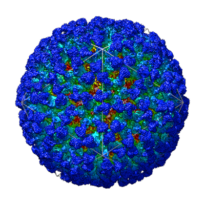 emd_22646.png
 http://ftp.pdbj.org/pub/emdb/structures/EMD-22646


 Z (Sec.)
 Y (Row.)
 X (Col.)


 Sample components
 Processing
 Electron microscopy
 FIELD EMISSION GUN
